
Search Count: 143
 |
Organism: Metagenome
Method: X-RAY DIFFRACTION Release Date: 2025-09-17 Classification: HYDROLASE Ligands: ACT, EDO, GOL, PEG, SO4 |
 |
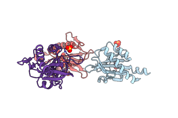
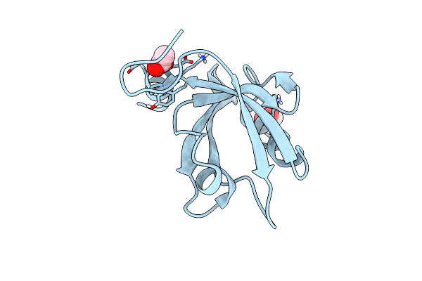
Structure Of The Ligand Binding Domain Of The Chemoreceptor Mkca (Dsomk10_Rs0100305) Of Dickeya Solani Mk10 In Complex With Choline
Organism: Dickeya solani mk10
Method: X-RAY DIFFRACTION Release Date: 2025-08-06 Classification: SIGNALING PROTEIN Ligands: CHT, GOL, EDO, SO4 |
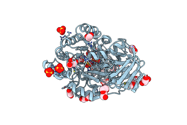 |
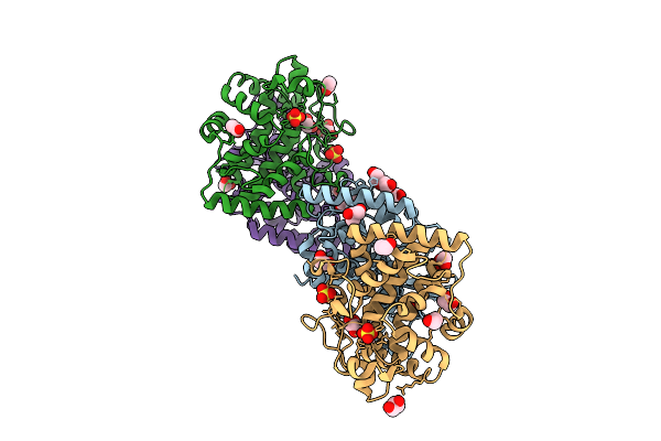
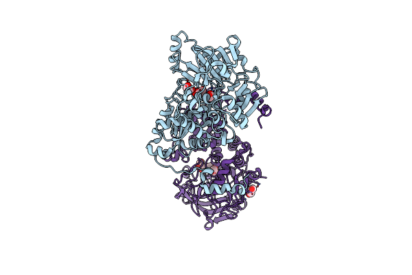
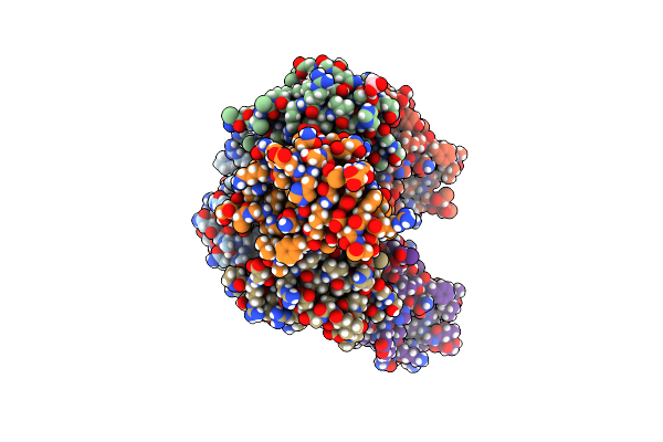
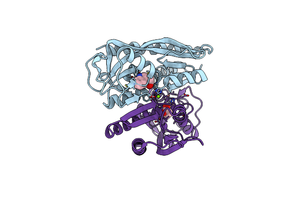
N-Acyl-D-Amino-Acid Deacylase (D-Acylase) From Klebsiella Pneumoniae In An Open Conformation
Organism: Klebsiella pneumoniae subsp. pneumoniae kp13
Method: X-RAY DIFFRACTION Release Date: 2025-06-18 Classification: HYDROLASE Ligands: SO4, PEG, EDO, GOL, PGE, NI |
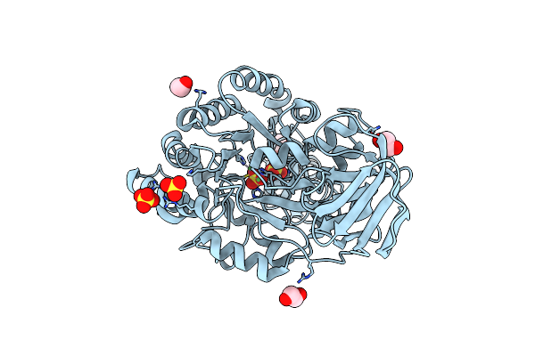 |
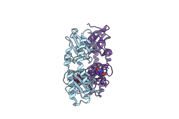
N-Acyl-D-Amino-Acid Deacylase (D-Acylase) From Klebsiella Pneumoniae In The Absence Of Glycerol
Organism: Klebsiella pneumoniae subsp. pneumoniae kp13
Method: X-RAY DIFFRACTION Release Date: 2025-06-18 Classification: HYDROLASE Ligands: EDO, SO4, NI |
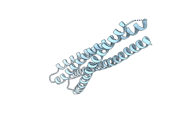 |
Single-Chain Chimeric Protein Mimicking The Interaction Between Hr1 And Hr2 In Hiv Gp41
Organism: Hiv-1 06tg.ht008
Method: X-RAY DIFFRACTION Resolution:2.30 Å Release Date: 2025-05-28 Classification: VIRAL PROTEIN |
 |
Organism: Synthetic construct
Method: X-RAY DIFFRACTION Resolution:2.10 Å Release Date: 2025-05-21 Classification: LYASE Ligands: GOL, EDO |
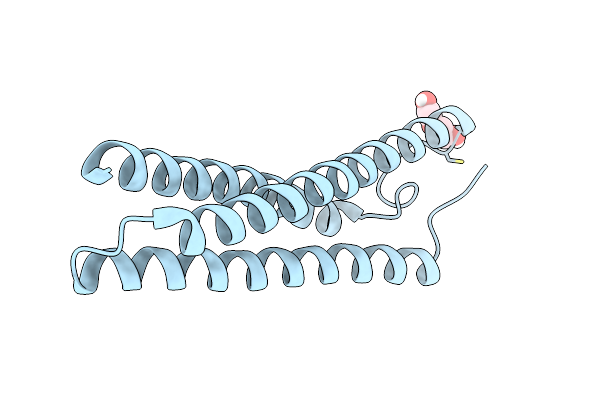 |
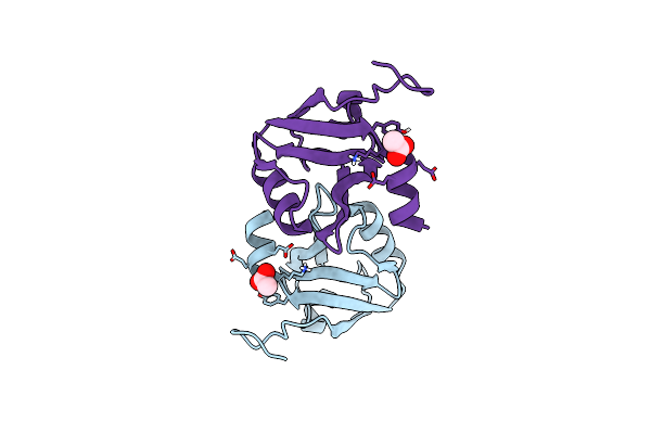
Anti-Hiv-1 Chimeric Miniprotein Mimicking The N-Terminal Half Of Gp41 Nhr With An Extended Region Targeting The Mper
Organism: Human immunodeficiency virus type 1 (bru isolate)
Method: X-RAY DIFFRACTION Resolution:1.70 Å Release Date: 2025-04-30 Classification: ANTIVIRAL PROTEIN Ligands: PEG |
 |
Sensor Domain Of Oscillibacter Ruminantium Chemoreceptor In Complex With Formate.
Organism: Oscillibacter ruminantium
Method: X-RAY DIFFRACTION Resolution:1.75 Å Release Date: 2025-02-12 Classification: SIGNALING PROTEIN Ligands: FMT, NA |
 |
Sensor Domain Of Asticcacaulis Benevestitus Chemoreceptor In Complex With Formate.
Organism: Asticcacaulis benevestitus
Method: X-RAY DIFFRACTION Resolution:2.10 Å Release Date: 2025-02-12 Classification: SIGNALING PROTEIN Ligands: FMT, SO4 |
 |
Ligand Binding Domain Of The P. Putida Receptor Mcph In Complex With Uric Acid
Organism: Pseudomonas putida kt2440
Method: X-RAY DIFFRACTION Resolution:1.95 Å Release Date: 2024-07-24 Classification: SIGNALING PROTEIN Ligands: URC |
 |
Organism: Homo sapiens
Method: X-RAY DIFFRACTION Resolution:2.70 Å Release Date: 2024-05-29 Classification: FLAVOPROTEIN Ligands: FAD, NAD |
 |
Organism: Homo sapiens
Method: X-RAY DIFFRACTION Resolution:2.50 Å Release Date: 2024-05-29 Classification: FLAVOPROTEIN Ligands: FAD, EPE |
 |
Structural Characterization Of Beta-Xyloxidase Xynb2 From Geobacillus Stearothermophilus Cect43
Organism: Geobacillus stearothermophilus
Method: X-RAY DIFFRACTION Resolution:2.25 Å Release Date: 2024-03-27 Classification: HYDROLASE Ligands: GOL, SO4, ACT |
 |
Organism: Escherichia coli
Method: X-RAY DIFFRACTION Release Date: 2023-10-18 Classification: HYDROLASE Ligands: EDO, SO4 |
 |
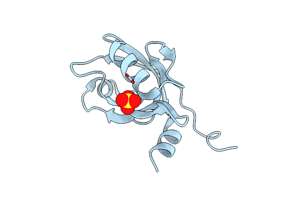
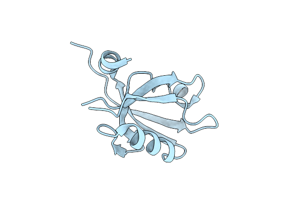
Crystal Structure Of The Third Pdz Domain Of Psd-95 Protein In The Space Group P3112 At Ph 4.0
Organism: Homo sapiens
Method: X-RAY DIFFRACTION Resolution:1.48 Å Release Date: 2023-03-08 Classification: PROTEIN BINDING Ligands: ACT |
 |
Crystal Structure Of The Third Pdz Domain Of Psd-95 Protein In The Space Group P212121 At Ph 4.6
Organism: Homo sapiens
Method: X-RAY DIFFRACTION Resolution:1.25 Å Release Date: 2023-03-08 Classification: PROTEIN BINDING Ligands: ACT |
 |
Crystal Structure Of The Third Pdz Domain Of Psd-95 Protein In The Space Group P21 At Ph 4.0
Organism: Homo sapiens
Method: X-RAY DIFFRACTION Resolution:1.63 Å Release Date: 2023-03-08 Classification: PROTEIN BINDING Ligands: ACT |
 |
Crystal Structure Of The Third Pdz Domain Of Psd-95 Protein In The Space Group P212121 At Ph 4.0
Organism: Homo sapiens
Method: X-RAY DIFFRACTION Resolution:1.25 Å Release Date: 2023-03-08 Classification: PROTEIN BINDING Ligands: SO4 |
 |
Crystal Structure Of The Third Pdz Domain Of Psd-95 Protein In The Space Group P3121 At Ph 3.7
Organism: Homo sapiens
Method: X-RAY DIFFRACTION Resolution:1.50 Å Release Date: 2023-03-08 Classification: PROTEIN BINDING |
 |
Structure Of The Ligand Binding Domain Of The Antibiotic Biosynthesis Regulator Admx From The Rhizobacterium Serratia Plymuthica A153 Bound To The Auxin Indole-3-Acetic Acid (Iaa).
Organism: Serratia plymuthica
Method: X-RAY DIFFRACTION Resolution:1.81 Å Release Date: 2022-12-14 Classification: TRANSCRIPTION Ligands: IAC, MG |