
Search Count: 27
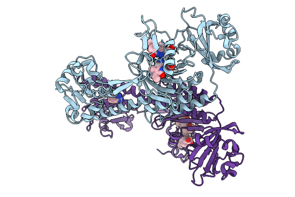 |
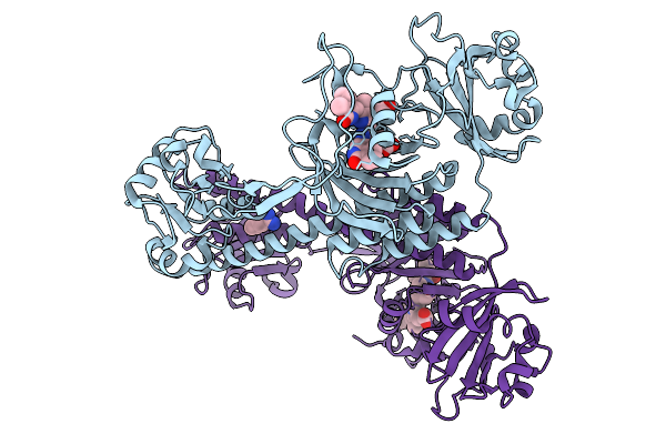
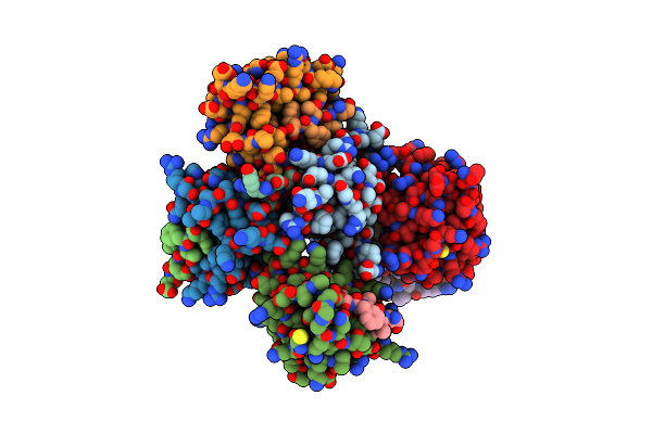
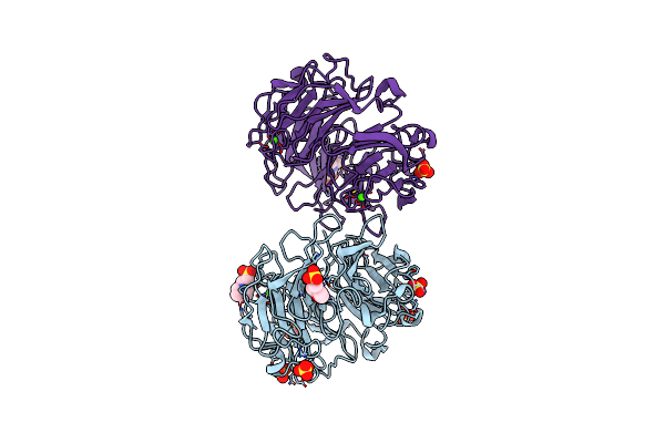
Photoactivation In Bacteriophytochromes, Reference (Dark) Structure For The 3 Ps Time Point
Organism: Stigmatella aurantiaca
Method: X-RAY DIFFRACTION Release Date: 2025-10-08 Classification: SIGNALING PROTEIN Ligands: 3Q8, BEN |
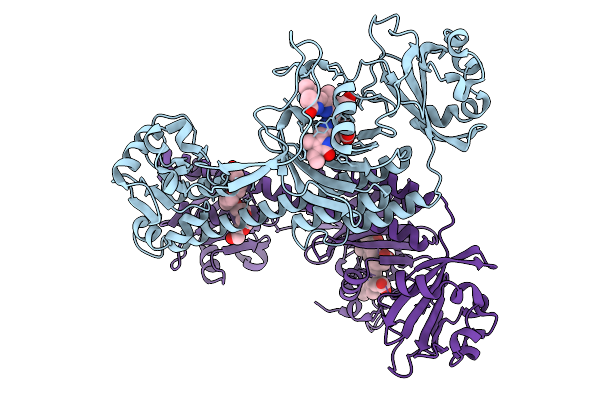 |
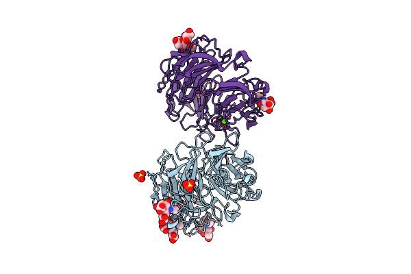
Photoactivation In Bacteriophytochrome, High Resolution Cryo Structure In The Dark.
Organism: Stigmatella aurantiaca
Method: X-RAY DIFFRACTION Release Date: 2025-10-08 Classification: SIGNALING PROTEIN Ligands: EL5, P33 |
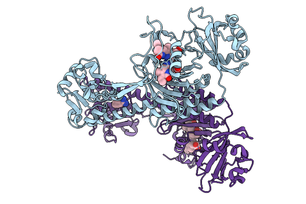 |
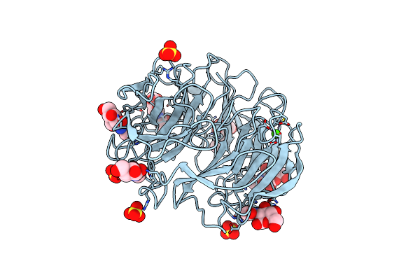
Photoactivation In Bacteriophytochromes, Reference (Dark) Structure For The 100 Ps Time Point
Organism: Stigmatella aurantiaca
Method: X-RAY DIFFRACTION Release Date: 2025-10-08 Classification: SIGNALING PROTEIN Ligands: EL5, BEN |
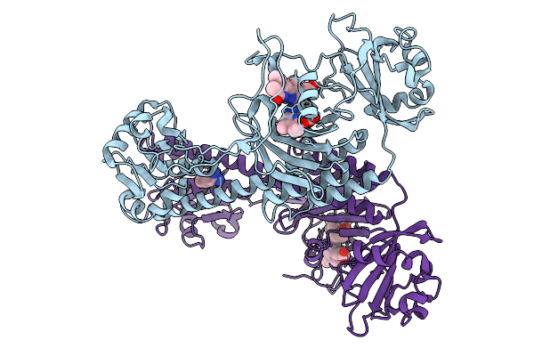 |
Organism: Stigmatella aurantiaca
Method: X-RAY DIFFRACTION Release Date: 2025-10-08 Classification: SIGNALING PROTEIN Ligands: BLA, BEN |
 |
Organism: Stigmatella aurantiaca
Method: X-RAY DIFFRACTION Release Date: 2025-10-08 Classification: SIGNALING PROTEIN Ligands: BLA, BEN |
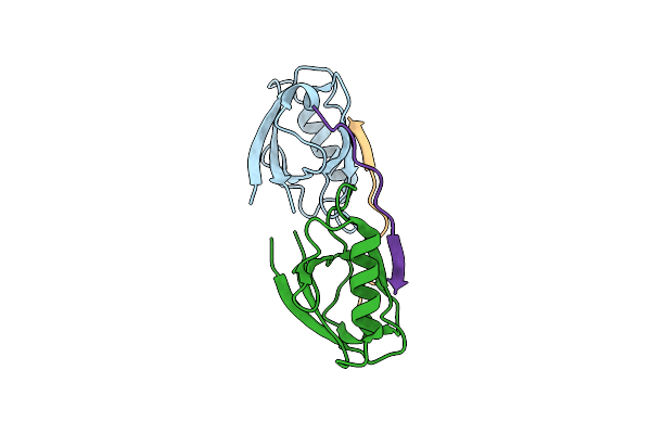 |
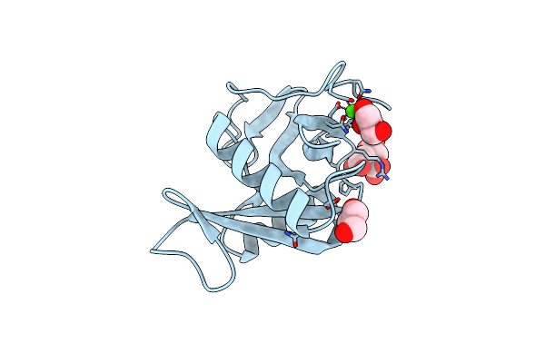
Crystal Structure Of Whirlin Pdz3 In Complex With Myosin 15A C-Terminal Pdz Binding Motif Peptide
Organism: Mus musculus
Method: X-RAY DIFFRACTION Resolution:1.70 Å Release Date: 2020-10-07 Classification: STRUCTURAL PROTEIN |
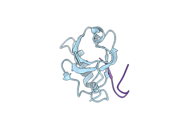 |
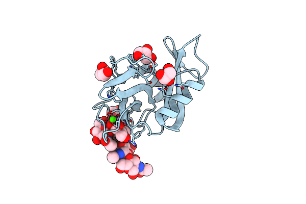
Crystal Structure Of Whirlin Pdz3_C-Ter In Complex With Myosin 15A C-Terminal Pdz Binding Motif Peptide
Organism: Mus musculus
Method: X-RAY DIFFRACTION Resolution:1.93 Å Release Date: 2020-10-07 Classification: STRUCTURAL PROTEIN |
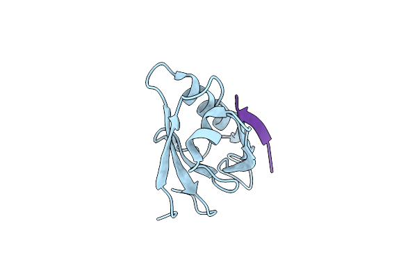 |
Crystal Structure Of Whirlin Pdz3_C-Ter In Complex With Cask Internal Pdz Binding Motif Peptide
Organism: Mus musculus
Method: X-RAY DIFFRACTION Resolution:1.63 Å Release Date: 2020-10-07 Classification: STRUCTURAL PROTEIN |
 |
Crystal Structure Of Whirlin Pdz3_C-Ter In Complex With Harmonin A1 C-Terminal Pdz Binding Motif Peptide
Organism: Mus musculus, Rattus norvegicus
Method: X-RAY DIFFRACTION Resolution:3.17 Å Release Date: 2020-10-07 Classification: STRUCTURAL PROTEIN |
 |
Crystal Structure Of Whirlin Pdz3_C-Ter In Complex With Taperin Internal Pdz Binding Motif Peptide
Organism: Mus musculus, Homo sapiens
Method: X-RAY DIFFRACTION Resolution:1.32 Å Release Date: 2020-10-07 Classification: STRUCTURAL PROTEIN |
 |
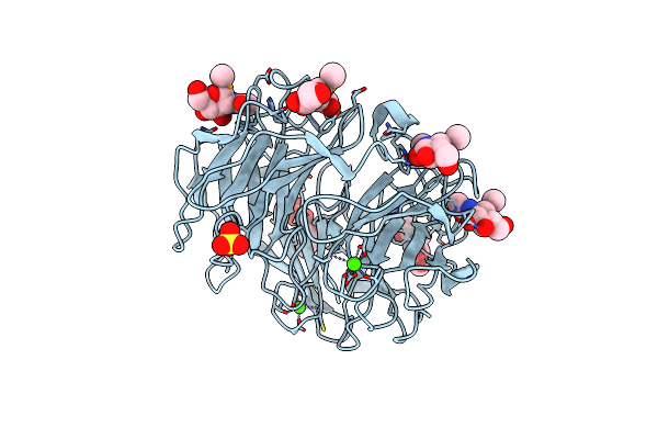
Organism: Codakia orbicularis
Method: X-RAY DIFFRACTION Resolution:1.30 Å Release Date: 2008-08-05 Classification: SUGAR BINDING PROTEIN Ligands: CA, GOL, CIT |
 |
Crystal Structure Of Codakine In Complex With Biantennary Nonasaccharide At 1.7A Resolution
Organism: Codakia orbicularis
Method: X-RAY DIFFRACTION Resolution:1.70 Å Release Date: 2008-08-05 Classification: SUGAR BINDING PROTEIN Ligands: CA, GOL |
 |
Crystal Structure Of Pseudomonas Aeruginosa Lectin (Pa-Iil) Complexed With Methyl-B-D-Arabinopyranoside
Organism: Pseudomonas aeruginosa
Method: X-RAY DIFFRACTION Resolution:1.80 Å Release Date: 2006-02-22 Classification: LECTIN Ligands: ARW, CA, SO4 |
 |
Crystal Structure Of Pseudomonas Aeruginosa Lectin (Pa-Iil) Complexed With A-L-Galactopyranoside
Organism: Pseudomonas aeruginosa
Method: X-RAY DIFFRACTION Resolution:1.50 Å Release Date: 2006-02-22 Classification: LECTIN Ligands: GXL, CA, SO4 |
 |
1.8A Crystal Structure Of Of Psathyrella Velutina Lectin In Complex With Methyl 2-Acetamido-1,2-Dideoxy-1-Seleno-Beta-D-Glucopyranoside
Organism: Psathyrella velutina
Method: X-RAY DIFFRACTION Resolution:1.80 Å Release Date: 2006-01-23 Classification: LECTIN Ligands: CA, SNG, SO4, GOL |
 |
Organism: Psathyrella velutina
Method: X-RAY DIFFRACTION Resolution:1.50 Å Release Date: 2006-01-23 Classification: LECTIN Ligands: CA, MES, CL, SO4 |
 |
1.8A Crystal Structure Of Psathyrella Velutina Lectin In Complex With N-Acetylneuraminic Acid
Organism: Psathyrella velutina
Method: X-RAY DIFFRACTION Resolution:1.80 Å Release Date: 2006-01-23 Classification: LECTIN Ligands: CA, SIA, SO4 |
 |
2.6A Crystal Structure Of Psathyrella Velutina Lectin In Complex With N-Acetylglucosamine
Organism: Psathyrella velutina
Method: X-RAY DIFFRACTION Resolution:2.60 Å Release Date: 2006-01-23 Classification: LECTIN Ligands: CA, NAG, SO4 |
 |
Pseudomonas Aeruginosa Lectin Ii (Pa-Iil)Complexed With Lacto-N-Neo- Fucopentaose V(Lnpfv)
Organism: Pseudomonas aeruginosa
Method: X-RAY DIFFRACTION Resolution:1.05 Å Release Date: 2005-03-31 Classification: SUGAR BINDING PROTEIN Ligands: SO4, CA, GOL |
 |
Structure Of Pseudomonas Aeruginosa Lectin Ii (Pa-Iil)Complexed With Lewisa Trisaccharide
Organism: Pseudomonas aeruginosa
Method: X-RAY DIFFRACTION Resolution:1.75 Å Release Date: 2005-03-31 Classification: SUGAR BINDING PROTEIN Ligands: CA, SO4, GOL |