
Search Count: 34
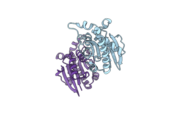 |
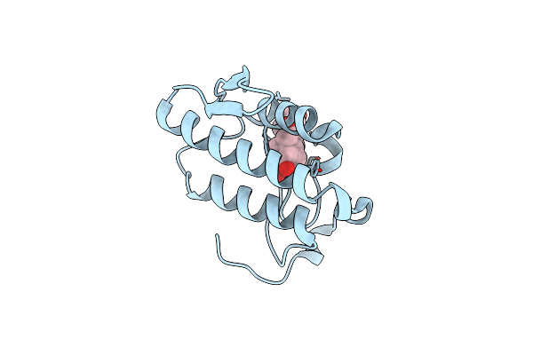
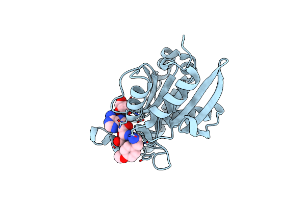
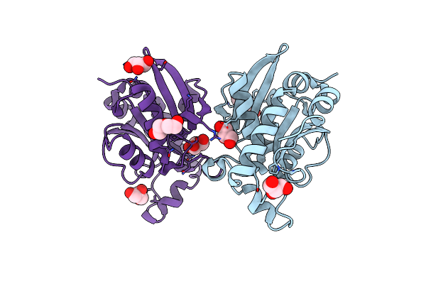
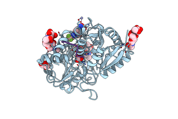
Crystal Structure Of Peptidyl-Trna Hydrolase From A Gram-Positive Bacterium, Streptococcus Pyogenes At 2.19 Angstrom Resolution Shows The Closed Structure Of The Substrate Binding Cleft
Organism: Streptococcus pyogenes nz131
Method: X-RAY DIFFRACTION Resolution:2.19 Å Release Date: 2014-08-06 Classification: HYDROLASE |
 |
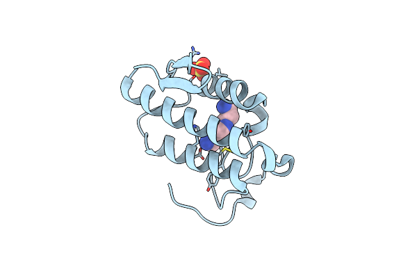
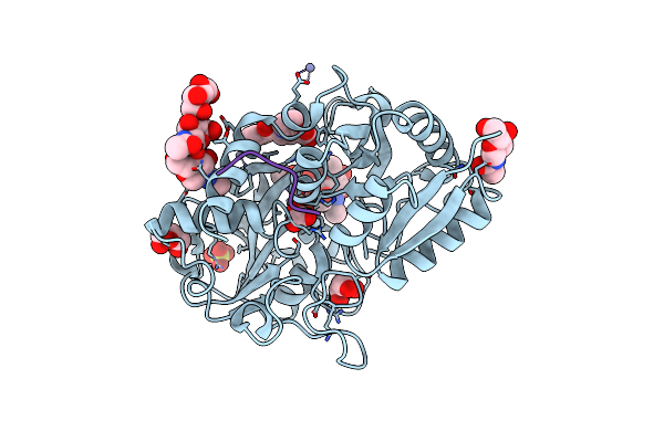
Crystal Structure Of Peptidyl-Trna Hydrolase From Pseudomonas Aeruginosa With 5-Azacytidine At 1.89 Angstrom Resolution
Organism: Pseudomonas aeruginosa
Method: X-RAY DIFFRACTION Resolution:1.89 Å Release Date: 2014-06-25 Classification: HYDROLASE Ligands: 5AE, GOL |
 |
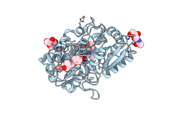
Crystal Structure Of The Complex Of Phospholipase A2 With P-Coumaric Acid At 1.2 A Resolution
Organism: Daboia russellii pulchella
Method: X-RAY DIFFRACTION Resolution:1.20 Å Release Date: 2014-06-18 Classification: HYDROLASE/HYDROLASE INHIBITOR Ligands: SO4, HC4 |
 |
Crystal Structure Of The Complex Of Phospholipase A2 With Resveratrol At 1.20 A Resolution
Organism: Daboia russellii pulchella
Method: X-RAY DIFFRACTION Resolution:1.20 Å Release Date: 2014-06-18 Classification: HYDROLASE/HYDROLASE INHIBITOR Ligands: SO4, STL |
 |
Crystal Structure Of The Complex Of Phospholipase A2 With Corticosterone At 1.48 A Resolution
Organism: Daboia russellii pulchella
Method: X-RAY DIFFRACTION Resolution:1.48 Å Release Date: 2014-06-18 Classification: HYDROLASE/HYDROLASE INHIBITOR Ligands: C0R |
 |
Crystal Structure Of The Complex Of Phospholipase A2 With Spermidine At 1.65 A Resolution
Organism: Daboia russellii pulchella
Method: X-RAY DIFFRACTION Resolution:1.65 Å Release Date: 2014-06-18 Classification: HYDROLASE/HYDROLASE INHIBITOR Ligands: SPD, SO4 |
 |
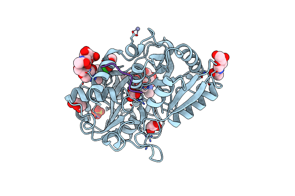
Crystal Structure Of The Complex Of Phospholipase A2 With Gramine Derivative At 1.80 A Resolution
Organism: Daboia russellii pulchella
Method: X-RAY DIFFRACTION Resolution:1.80 Å Release Date: 2014-06-18 Classification: HYDROLASE/HYDROLASE INHIBITOR Ligands: PZZ |
 |
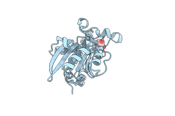
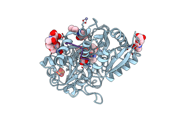
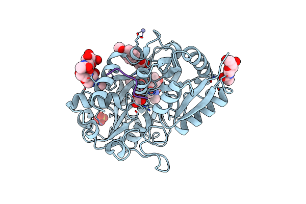
Crystal Structure Of Peptidyl-Trna Hydrolase From Pseudomonas Aeruginosa At 1.5 Angstrom Resolution
Organism: Pseudomonas aeruginosa
Method: X-RAY DIFFRACTION Resolution:1.50 Å Release Date: 2014-05-28 Classification: HYDROLASE |
 |
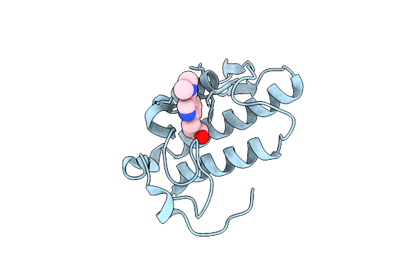
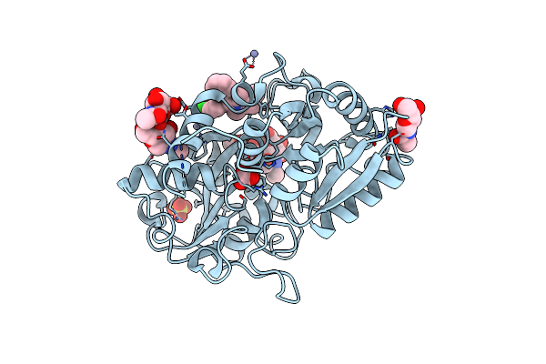
Crystal Structure Of The Complex Of Peptidyl-Trna Hydrolase From Pseudomonas Aeruginosa With Amino Acyl-Trna Analogue At 1.77 Angstrom Resolution
Organism: Pseudomonas aeruginosa
Method: X-RAY DIFFRACTION Resolution:1.77 Å Release Date: 2014-05-28 Classification: HYDROLASE Ligands: GOL, 3NZ |
 |
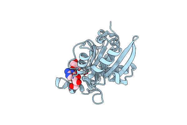
Crystal Structure Of C-Lobe Of Bovine Lactoferrin Complexed With Fenoprofen At 2.1 Angstrom Resolution
Organism: Bos taurus
Method: X-RAY DIFFRACTION Resolution:2.10 Å Release Date: 2013-12-11 Classification: HYDROLASE Ligands: ZN, FE, CO3, SO4, NAG, PFN |
 |
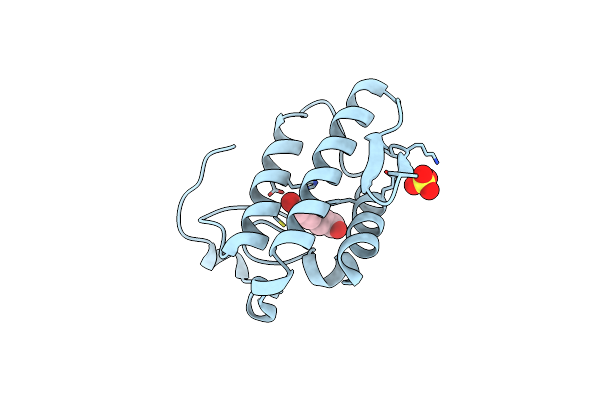
Crystal Structure Of C-Lobe Of Bovine Lactoferrin Complexed With Meclofenamic Acid At 1.4 A Resolution
Organism: Bos taurus
Method: X-RAY DIFFRACTION Resolution:1.40 Å Release Date: 2013-10-30 Classification: HYDROLASE Ligands: ZN, FE, CO3, SO4, NAG, GOL, JMS |
 |
Crystal Structure Of Peptidyl-Trna Hydrolase From Pseudomonas Aeruginosa At 2.25 Angstrom Resolution
Organism: Pseudomonas aeruginosa
Method: X-RAY DIFFRACTION Resolution:2.25 Å Release Date: 2013-04-03 Classification: HYDROLASE Ligands: GOL |
 |
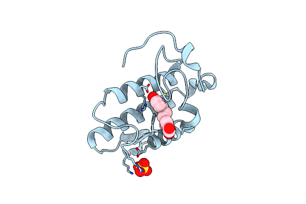
Crystal Structure Of C-Lobe Of Bovine Lactoferrin Complexed With Ketorolac At 1.68 A Resolution
Organism: Bos taurus
Method: X-RAY DIFFRACTION Resolution:1.68 Å Release Date: 2012-09-19 Classification: HYDROLASE Ligands: NAG, ZN, FE, CO3, SO4, GOL, KTR |
 |
Crystal Structure Of C-Lobe Of Bovine Lactoferrin Complexed With Tolfenamic Acid At 1.98 A Resolution
Organism: Bos taurus
Method: X-RAY DIFFRACTION Resolution:1.98 Å Release Date: 2012-08-29 Classification: HYDROLASE Ligands: FE, CO3, SO4, ZN, NAG, TLF |
 |
Crystal Structure Of C-Lobe Of Bovine Lactoferrin Complexed With Licofelone At 1.88 A Resolution
Organism: Bos taurus
Method: X-RAY DIFFRACTION Resolution:1.88 Å Release Date: 2012-08-29 Classification: HYDROLASE Ligands: NAG, ZN, FE, CO3, SO4, LCF |
 |
Crystal Structure Of C-Lobe Of Bovine Lactoferrin Complexed With Mefenamic Acid At 1.90 A Resolution
Organism: Bos taurus
Method: X-RAY DIFFRACTION Resolution:1.90 Å Release Date: 2012-08-01 Classification: METAL BINDING PROTEIN Ligands: NAG, FE, ZN, CO3, SO4, ID8 |
 |
Crystal Structure Of Peptidyl T-Rna Hydrolase From Pseudomonas Aeruginosa At 2.2 Angstrom Resolution
Organism: Pseudomonas aeruginosa
Method: X-RAY DIFFRACTION Resolution:2.25 Å Release Date: 2012-07-04 Classification: HYDROLASE Ligands: PEG, GOL |
 |
Crystal Structure Of C-Lobe Of Bovine Lactoferrin Complexed With Flurbiprofen At 1.58 A Resolution
Organism: Bos taurus
Method: X-RAY DIFFRACTION Resolution:1.58 Å Release Date: 2012-07-04 Classification: HYDROLASE Ligands: FE, CO3, SO4, ZN, NAG, FLP, GOL |
 |
Crystal Structure Of C-Lobe Of Bovine Lactoferrin Complexed With Celecoxib Acid At 1.80 A Resolution
Organism: Bos taurus
Method: X-RAY DIFFRACTION Resolution:1.80 Å Release Date: 2012-06-27 Classification: HYDROLASE Ligands: FE, CO3, ZN, CEL, GOL |
 |
Crystal Structure Of C-Lobe Of Bovine Lactoferrin Complexed With Naproxen At 1.68 A Resolution
Organism: Bos taurus
Method: X-RAY DIFFRACTION Resolution:1.68 Å Release Date: 2012-06-27 Classification: HYDROLASE Ligands: NAG, FE, ZN, CO3, SO4, NPS |