
Search Count: 18
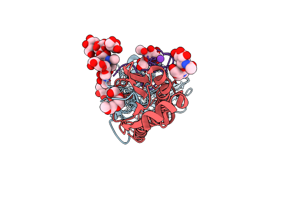 |
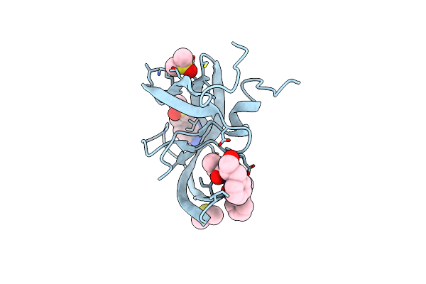
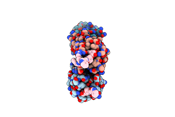
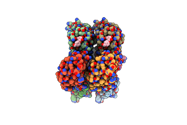
Crystal Structure Of The A/Puerto Rico/8/1934 (H1N1) Influenza Virus Hemagglutinin In Complex With D-Peptide
Organism: Influenza a virus (a/puerto rico/8/1934(h1n1)), Phage display vector pdvec0
Method: X-RAY DIFFRACTION Release Date: 2025-06-25 Classification: VIRAL PROTEIN Ligands: NAG, TOE, K |
 |
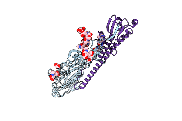
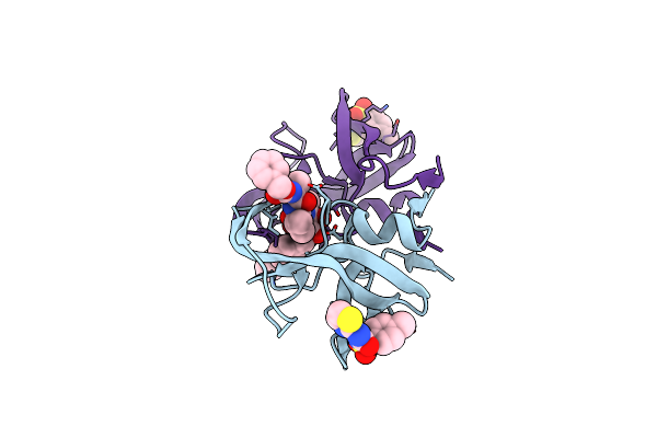
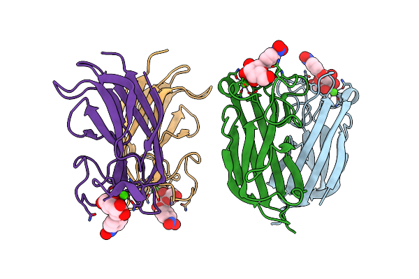
Crystal Structure Of Puf60 Uhm Domain In Complex With 7,8 Dimethoxyperphenazine
Organism: Citrobacter freundii mgh 56, Homo sapiens
Method: X-RAY DIFFRACTION Resolution:1.94 Å Release Date: 2020-09-09 Classification: SPLICING Ligands: MG, LJT |
 |
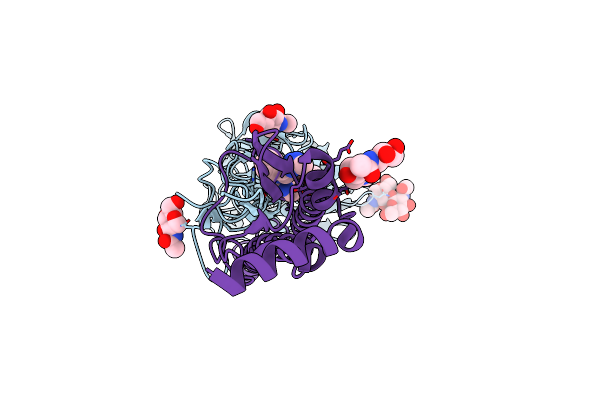
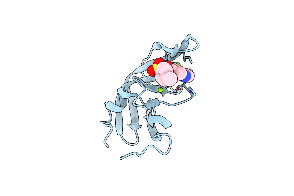
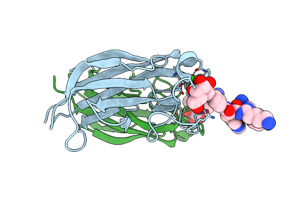
Crystal Structure Of The A/Solomon Islands/3/2006(H1N1) Influenza Virus Hemagglutinin In Complex With Small Molecule Jnj4796
Organism: Influenza a virus, Influenza a virus (a/solomon islands/3/2006(h1n1))
Method: X-RAY DIFFRACTION Resolution:2.72 Å Release Date: 2019-02-13 Classification: VIRAL PROTEIN Ligands: NAG, EZ7 |
 |
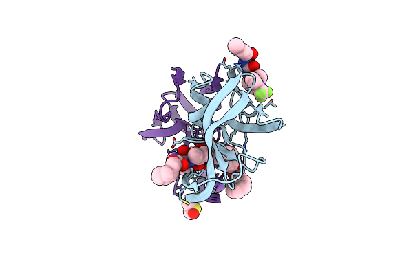
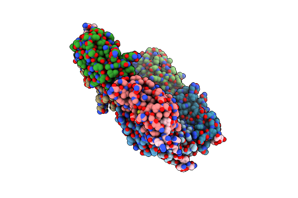
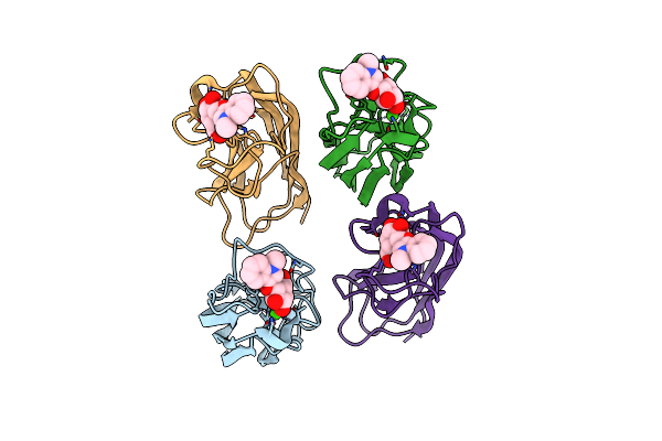
Crystal Structure Of The A/Vietnam/1203/2004 (H5N1) Influenza Virus Hemagglutinin In Complex With Small Molecule Jnj4796
Organism: Influenza a virus (a/viet nam/1203/2004(h5n1))
Method: X-RAY DIFFRACTION Resolution:2.32 Å Release Date: 2019-02-13 Classification: VIRAL PROTEIN Ligands: NAG, EZ7, PEG |
 |
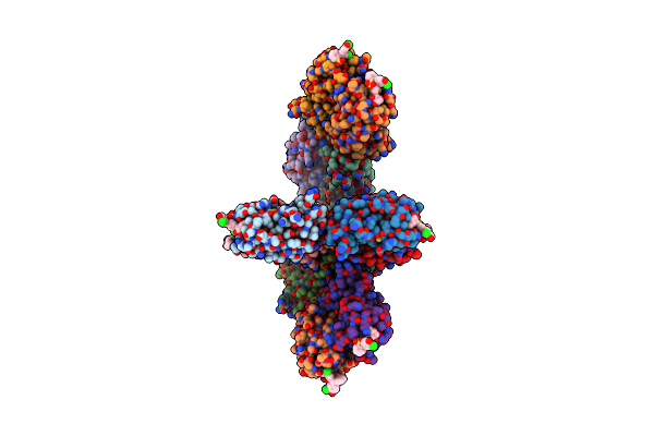
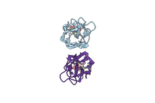
Hiv Protease (Pr) In Open Form With Mg2+ In Active Site And Hive-9 In Eye Site
Organism: Human immunodeficiency virus 1
Method: X-RAY DIFFRACTION Resolution:3.00 Å Release Date: 2018-06-20 Classification: HYDROLASE Ligands: 3TL, HV9, DMS |
 |
Hiv Protease (Pr) With Tl-3 In Active Site And 4-Methylbenzene-1,2-Diamine In Exosite
Organism: Human immunodeficiency virus 1
Method: X-RAY DIFFRACTION Resolution:2.00 Å Release Date: 2018-04-25 Classification: HYDROLASE Ligands: 3TL, 9AY, DMS |
 |
Hiv Protease (Pr) With Tl-3 In The Active Site And (Z)-N-(Thiazol-2-Yl)-N'-Tosylcarbamimidate In The Exosite
Organism: Human immunodeficiency virus 1
Method: X-RAY DIFFRACTION Resolution:1.80 Å Release Date: 2018-04-18 Classification: HYDROLASE Ligands: 3TL, 7GC |
 |
Hiv Protease (Pr) In Open Form With Mg2+ In Active Site And Hive-9 In Eye Site
Organism: Human immunodeficiency virus 1
Method: X-RAY DIFFRACTION Resolution:2.00 Å Release Date: 2018-04-18 Classification: HYDROLASE Ligands: HV9, MG |
 |
Organism: Homo sapiens
Method: X-RAY DIFFRACTION Resolution:2.22 Å Release Date: 2016-10-26 Classification: Splicing/Inhibitor |
 |
Crystal Structure Of Pseudomonas Aeruginosa Lectin Leca Complexed With Gala-Qrs At 2.31 A Resolution
Organism: Pseudomonas aeruginosa
Method: X-RAY DIFFRACTION Resolution:2.31 Å Release Date: 2013-12-18 Classification: SUGAR BINDING PROTEIN/INHIBITOR Ligands: GAL, CA, PHB |
 |
Crystal Structure Of Pseudomonas Aeruginosa Lectin Leca Complexed With Gala-Wri At 1.65 A Resolution
Organism: Pseudomonas aeruginosa
Method: X-RAY DIFFRACTION Resolution:1.65 Å Release Date: 2013-12-18 Classification: SUGAR BINDING PROTEIN/INHIBITOR Ligands: GAL, CA, PHB |
 |
Crystal Structure Of Pseudomonas Aeruginosa Lectin Leca Complexed With Gala-Wky At 1.64 A Resolution
Organism: Pseudomonas aeruginosa
Method: X-RAY DIFFRACTION Resolution:1.64 Å Release Date: 2013-12-18 Classification: SUGAR BINDING PROTEIN/INHIBITOR Ligands: GAL, CA, PHB |
 |
Crystal Structure Of Pseudomonas Aeruginosa Lectin Leca Complexed With 1-Methyl-3-Indolyl-B-D-Galactopyranoside At 1.45 A Resolution
Organism: Pseudomonas aeruginosa
Method: X-RAY DIFFRACTION Resolution:1.45 Å Release Date: 2013-10-30 Classification: SUGAR BINDING PROTEIN Ligands: CA, MHD, GAL |
 |
Crystal Structure Of Pseudomonas Aeruginosa Lectin Leca Complexed With Chlorophenol Red-B-D-Galactopyranoside At 2.86 A Resolution
Organism: Pseudomonas aeruginosa
Method: X-RAY DIFFRACTION Resolution:2.86 Å Release Date: 2013-10-30 Classification: SUGAR BINDING PROTEIN Ligands: CA, LRD, GAL |
 |
Crystal Structure Of Pseudomonas Aeruginosa Lectin Leca Complexed With Resorufin-B-D-Galactopyranoside At 1.76 A Resolution
Organism: Pseudomonas aeruginosa
Method: X-RAY DIFFRACTION Resolution:1.76 Å Release Date: 2013-10-30 Classification: SUGAR BINDING PROTEIN Ligands: CA, 04G, GAL |
 |
Crystal Structure Of Pa-Il Lectin Complexed With Galag0 At 2.3 A Resolution
Organism: Pseudomonas aeruginosa (strain atcc 15692 / dsm 22644 / cip 104116 / jcm 14847 / lmg 12228 / 1c / prs 101 / pao1), Synthetic construct
Method: X-RAY DIFFRACTION Resolution:2.29 Å Release Date: 2011-09-21 Classification: SUGAR BINDING PROTEIN Ligands: CA, GAL, PHB |
 |
Organism: Pseudomonas aeruginosa
Method: X-RAY DIFFRACTION Resolution:1.94 Å Release Date: 2011-09-21 Classification: SUGAR BINDING PROTEIN Ligands: CA, 147 |
 |
Crystal Structure Of Pa-Il Lectin Complexed With Galbg0 At 1.5 A Resolution
Organism: Pseudomonas aeruginosa (strain atcc 15692 / dsm 22644 / cip 104116 / jcm 14847 / lmg 12228 / 1c / prs 101 / pao1), Synthetic construct
Method: X-RAY DIFFRACTION Resolution:1.50 Å Release Date: 2011-09-21 Classification: SUGAR BINDING PROTEIN/INHIBITOR Ligands: CA, G0S |