
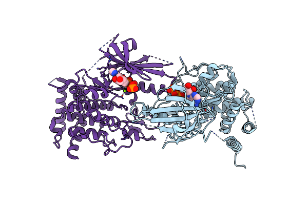
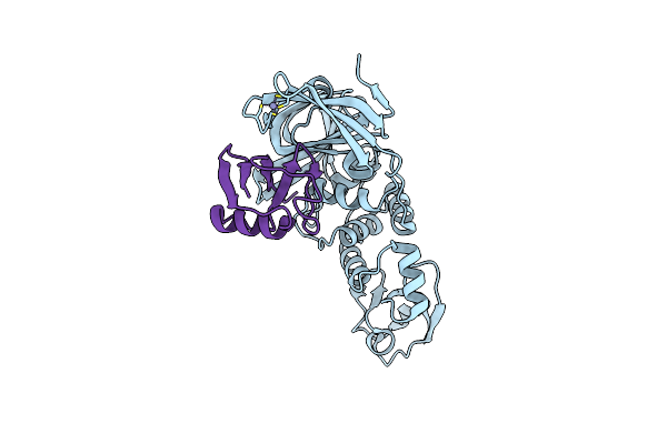
Search Count: 21
 |
Organism: Homo sapiens
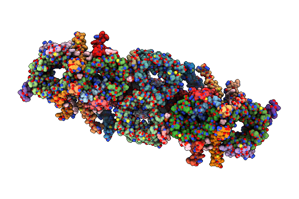
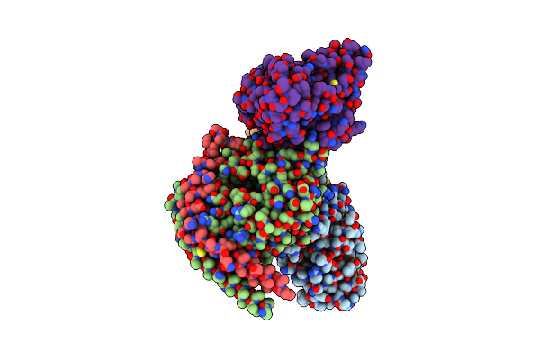
Method: ELECTRON MICROSCOPY Resolution:3.10 Å Release Date: 2025-03-12 Classification: TRANSLOCASE Ligands: PC1 |
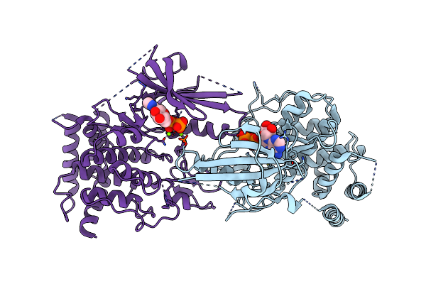
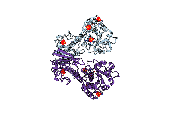
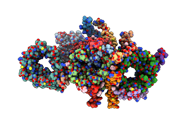 |
Organism: Homo sapiens
Method: ELECTRON MICROSCOPY Resolution:2.75 Å Release Date: 2025-03-12 Classification: TRANSLOCASE Ligands: PC1 |
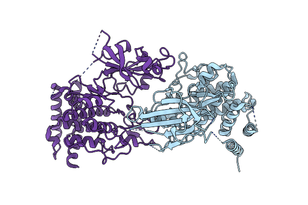
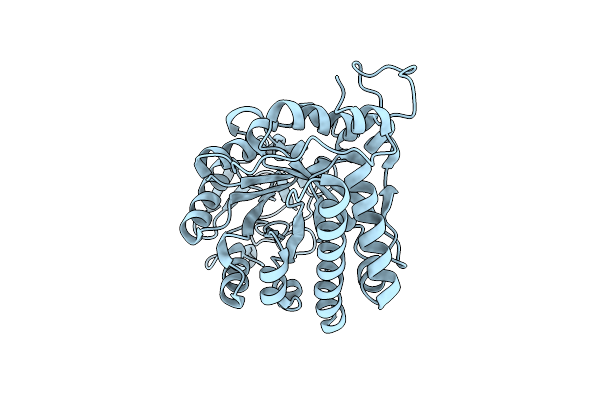
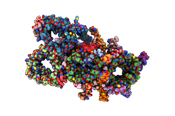 |
Organism: Homo sapiens
Method: ELECTRON MICROSCOPY Resolution:3.30 Å Release Date: 2025-03-12 Classification: TRANSLOCASE Ligands: PC1 |
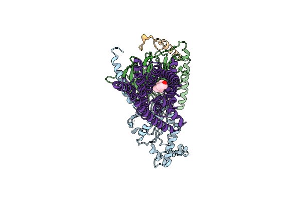 |
Identification, Structure And Agonist Design Of An Androgen Membrane Receptor.
Organism: Rattus norvegicus, Bos taurus, Homo sapiens
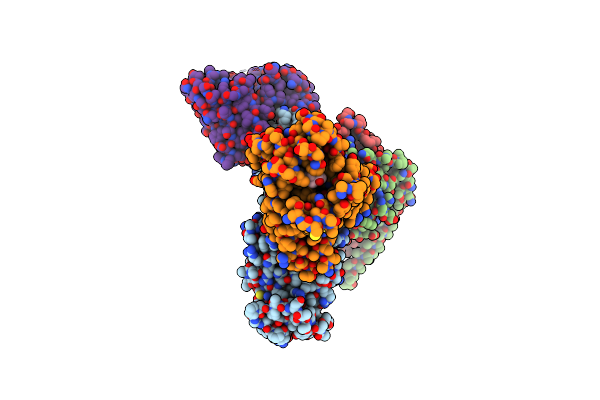
Method: ELECTRON MICROSCOPY Release Date: 2025-02-12 Classification: MEMBRANE PROTEIN Ligands: DHT |
 |
Identification, Structure And Agonist Design Of An Androgen Membrane Receptor
Organism: Bos taurus, Rattus norvegicus, Homo sapiens, Synthetic construct
Method: ELECTRON MICROSCOPY Release Date: 2025-02-12 Classification: MEMBRANE PROTEIN Ligands: YNH |
 |
Identification, Structure And Agonist Design Of An Androgen Membrane Receptor
Organism: Rattus norvegicus, Bos taurus, Synthetic construct, Homo sapiens
Method: ELECTRON MICROSCOPY Release Date: 2025-02-12 Classification: MEMBRANE PROTEIN Ligands: A1LU1 |
 |
Identification, Structure And Agonist Design Of An Androgen Membrane Receptor.
Organism: Synthetic construct, Homo sapiens
Method: ELECTRON MICROSCOPY Release Date: 2025-02-12 Classification: MEMBRANE PROTEIN Ligands: DHT |
 |
Identification, Structure And Agonist Design Of An Androgen Membrane Receptor.
Organism: Homo sapiens
Method: ELECTRON MICROSCOPY Release Date: 2025-02-12 Classification: MEMBRANE PROTEIN Ligands: DHT |
 |
Organism: Pediculus humanus corporis
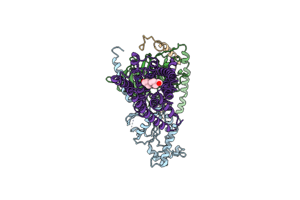
Method: ELECTRON MICROSCOPY Release Date: 2024-01-31 Classification: TRANSFERASE |
 |
Organism: Pediculus humanus corporis
Method: ELECTRON MICROSCOPY Release Date: 2024-01-31 Classification: TRANSFERASE Ligands: ANP, MG |
 |
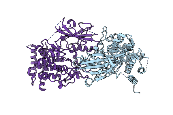
Structure Of Adp-Bound And Phosphorylated Pediculus Humanus (Ph) Pink1 Dimer
Organism: Pediculus humanus corporis
Method: ELECTRON MICROSCOPY Release Date: 2024-01-31 Classification: TRANSFERASE Ligands: ADP, MG |
 |
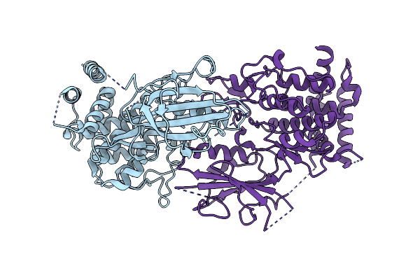
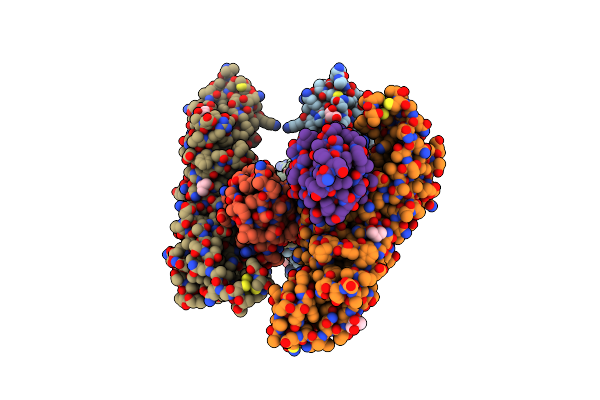
Structure Of Dimeric Phosphorylated Pediculus Humanus (Ph) Pink1 With Kinked Alpha-C Helix In Chain B
Organism: Pediculus humanus corporis
Method: ELECTRON MICROSCOPY Release Date: 2022-01-12 Classification: TRANSFERASE |
 |
Structure Of Dimeric Phosphorylated Pediculus Humanus (Ph) Pink1 With Extended Alpha-C Helix In Chain B
Organism: Pediculus humanus corporis
Method: ELECTRON MICROSCOPY Release Date: 2022-01-12 Classification: TRANSFERASE |
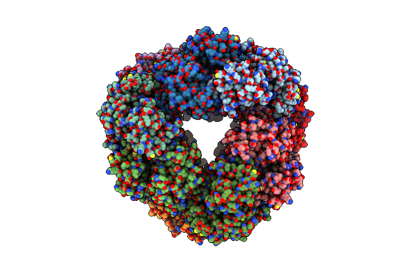 |
Structure Of Dodecameric Unphosphorylated Pediculus Humanus (Ph) Pink1 D357A Mutant
Organism: Pediculus humanus corporis
Method: ELECTRON MICROSCOPY Release Date: 2022-01-12 Classification: TRANSFERASE |
 |
Structure Of Dimeric Unphosphorylated Pediculus Humanus (Ph) Pink1 D357A Mutant
Organism: Pediculus humanus corporis
Method: ELECTRON MICROSCOPY Release Date: 2022-01-12 Classification: TRANSFERASE |
 |
Organism: Pediculus humanus corporis
Method: X-RAY DIFFRACTION Resolution:3.53 Å Release Date: 2021-12-22 Classification: TRANSFERASE |
 |
Organism: Severe acute respiratory syndrome coronavirus 2, Homo sapiens
Method: X-RAY DIFFRACTION Resolution:2.90 Å Release Date: 2020-06-17 Classification: HYDROLASE/SUBSTRATE Ligands: GOL, ZN |
 |
Organism: Severe acute respiratory syndrome coronavirus 2, Homo sapiens
Method: X-RAY DIFFRACTION Resolution:2.70 Å Release Date: 2020-06-17 Classification: HYDROLASE/SUBSTRATE Ligands: ZN |
 |
Crystal Structure Of Ribbx, A Two Domain 3,4-Dihydroxy-2-Butanone 4-Phosphate Synthase From A. Baumannii.
Organism: Acinetobacter baumannii
Method: X-RAY DIFFRACTION Resolution:2.66 Å Release Date: 2019-04-17 Classification: LYASE Ligands: SO4, CL |
 |
The Crystal Structure Of A Chitinase Crchi1 From The Nematophagous Fungus Clonostachys Rosea
Organism: Bionectria ochroleuca
Method: X-RAY DIFFRACTION Resolution:1.80 Å Release Date: 2010-02-16 Classification: HYDROLASE |