
Search Count: 111
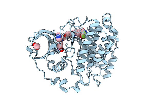 |
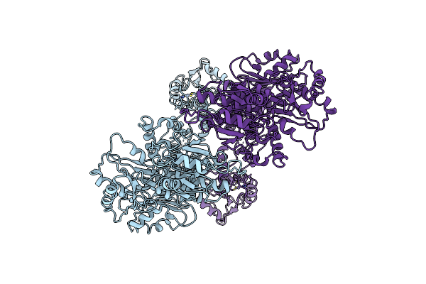
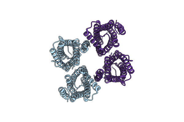
Organism: Homo sapiens
Method: X-RAY DIFFRACTION Release Date: 2025-10-22 Classification: HYDROLASE Ligands: A1I9L, ACT |
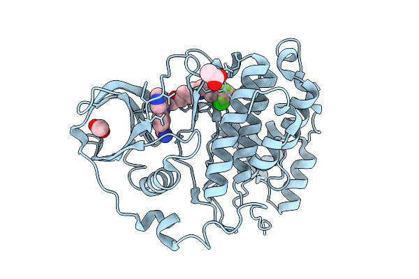 |
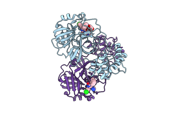
Organism: Homo sapiens
Method: X-RAY DIFFRACTION Release Date: 2025-10-22 Classification: HYDROLASE Ligands: A1JB4, ACT |
 |
Crystal Structure Of Sars-Cov-2 Main Protease (Mpro)In Complex With Inhibitor Avi-3318
Organism: Severe acute respiratory syndrome coronavirus 2
Method: X-RAY DIFFRACTION Release Date: 2025-07-23 Classification: HYDROLASE/HYDROLASE INHIBITOR Ligands: A1BTM |
 |
Crystal Structure Of Sars-Cov-2 Main Protease (Mpro) In Complex With Inhibitor Avi-4692
Organism: Severe acute respiratory syndrome coronavirus 2
Method: X-RAY DIFFRACTION Release Date: 2025-07-23 Classification: HYDROLASE Ligands: A1BVT, A1BTO |
 |
Crystal Structure Of Sars-Cov-2 Main Protease (Mpro)In Complex With Inhibitor Avi-4516
Organism: Severe acute respiratory syndrome coronavirus 2
Method: X-RAY DIFFRACTION Release Date: 2025-07-23 Classification: HYDROLASE/HYDROLASE INHIBITOR Ligands: A1BTP, EDO |
 |
Crystal Structure Of Sars-Cov-2 Main Protease (Mpro) Variant Q192T In Complex With Inhibitor Avi-4303
Organism: Severe acute respiratory syndrome coronavirus 2
Method: X-RAY DIFFRACTION Release Date: 2025-07-23 Classification: HYDROLASE/HYDROLASE INHIBITOR Ligands: EDO, A1BTN, CL |
 |
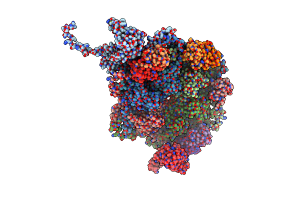
Organism: Homo sapiens
Method: ELECTRON MICROSCOPY Release Date: 2025-06-18 Classification: HYDROLASE Ligands: ZN |
 |
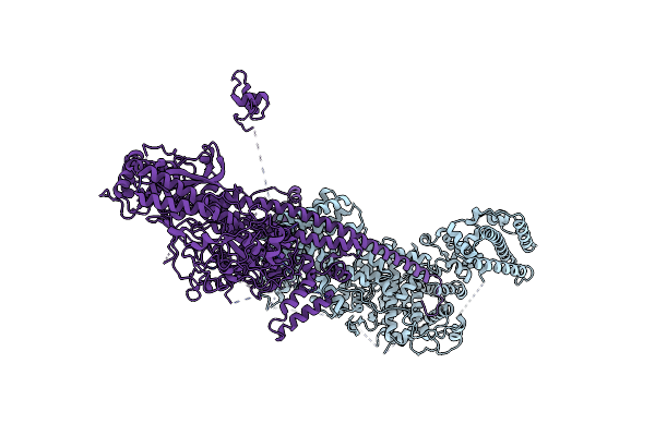
Cryo-Em Structure Of Human Dynactin Complex Bound To Chlamydia Effector Dre1
Organism: Homo sapiens
Method: ELECTRON MICROSCOPY Release Date: 2025-04-16 Classification: MOTOR PROTEIN Ligands: ADP, ANP, ZN |
 |
Cryo-Em Structure Of Human Dynactin Complex Bound To Chlamydia Effector Dre1
Organism: Homo sapiens
Method: ELECTRON MICROSCOPY Release Date: 2025-04-16 Classification: MOTOR PROTEIN Ligands: ADP, ANP, ZN |
 |
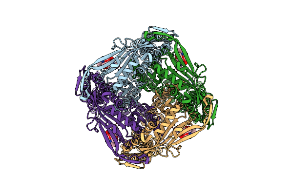
Organism: Anabaena phage a-4l
Method: ELECTRON MICROSCOPY Release Date: 2025-04-09 Classification: VIRUS |
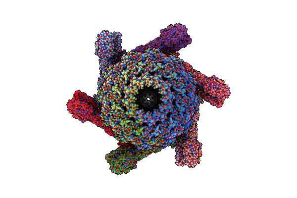 |
Organism: Anabaena phage a-4l
Method: ELECTRON MICROSCOPY Release Date: 2025-04-09 Classification: VIRAL PROTEIN |
 |
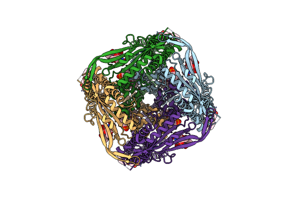
Organism: Anabaena phage a-4l
Method: ELECTRON MICROSCOPY Release Date: 2025-04-09 Classification: VIRAL PROTEIN Ligands: ZN |
 |
Organism: Anabaena phage a-4l
Method: ELECTRON MICROSCOPY Release Date: 2025-04-09 Classification: VIRAL PROTEIN |
 |
Cryo-Em Structure Of Mouse Trpml1 Channel Y404W At 2.86 Angstrom Resolution
Organism: Mus musculus
Method: ELECTRON MICROSCOPY Release Date: 2024-10-09 Classification: MEMBRANE PROTEIN |
 |
Cryo-Em Structure Of Mouse Pi(4,5)P2-Bound Trpml1 Channel At 2.46 Angstrom Resolution
Organism: Mus musculus
Method: ELECTRON MICROSCOPY Release Date: 2024-10-09 Classification: MEMBRANE PROTEIN Ligands: PIO, FO4 |
 |
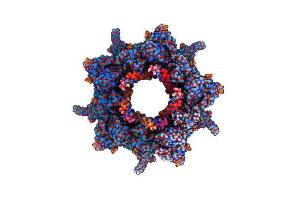
Organism: Caenorhabditis elegans
Method: ELECTRON MICROSCOPY Release Date: 2024-08-21 Classification: MEMBRANE PROTEIN |
 |
Organism: Caenorhabditis elegans
Method: ELECTRON MICROSCOPY Release Date: 2024-08-21 Classification: MEMBRANE PROTEIN |
 |
Organism: Mus musculus
Method: ELECTRON MICROSCOPY Release Date: 2024-08-21 Classification: MEMBRANE PROTEIN |
 |
Organism: Mus musculus
Method: ELECTRON MICROSCOPY Release Date: 2024-08-21 Classification: MEMBRANE PROTEIN |
 |
Organism: Mus musculus
Method: ELECTRON MICROSCOPY Release Date: 2024-08-21 Classification: MEMBRANE PROTEIN |