Search Count: 27
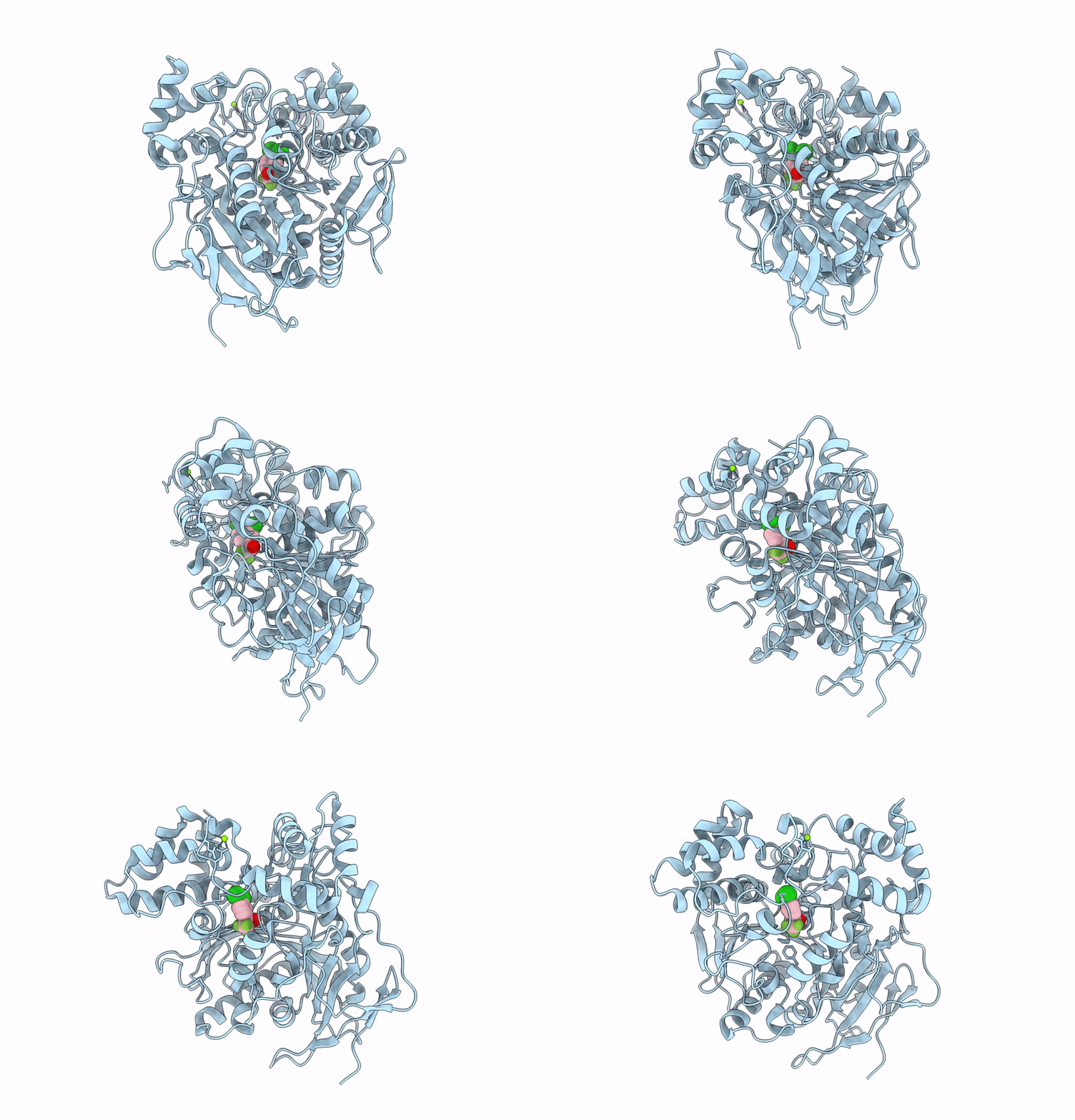 |
Organism: Homo sapiens
Method: X-RAY DIFFRACTION Resolution:1.83 Å Release Date: 2025-01-29 Classification: HYDROLASE Ligands: A1EIY, MG |
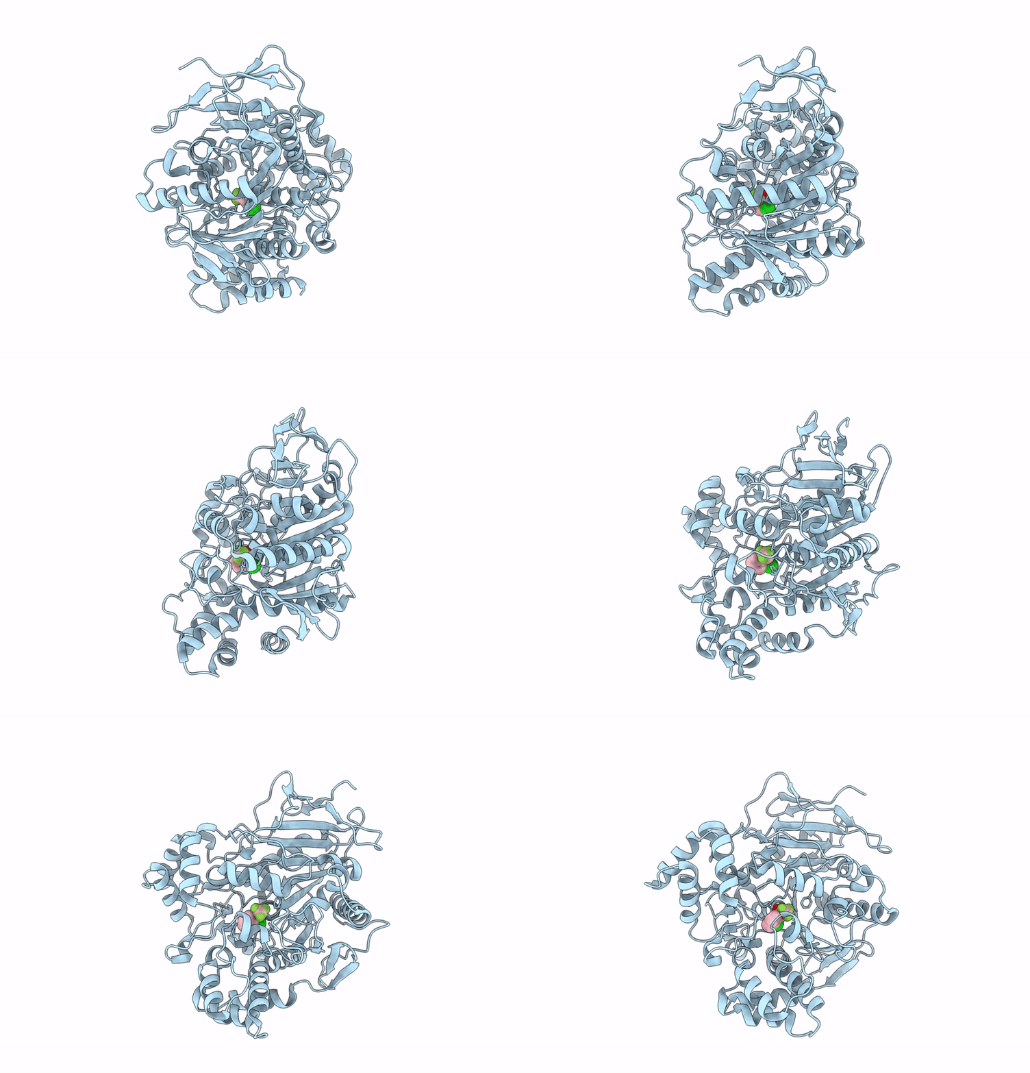 |
Organism: Homo sapiens
Method: X-RAY DIFFRACTION Resolution:1.89 Å Release Date: 2025-01-29 Classification: HYDROLASE Ligands: A1EIZ |
 |
Organism: Sus scrofa
Method: ELECTRON MICROSCOPY Release Date: 2023-08-30 Classification: MEMBRANE PROTEIN Ligands: PCW, MG, PWI, NAG, CLR |
 |
Organism: Sus scrofa
Method: ELECTRON MICROSCOPY Release Date: 2023-08-30 Classification: MEMBRANE PROTEIN Ligands: PCW, MG, PXR, NAG, CLR |
 |
Organism: Sus scrofa
Method: ELECTRON MICROSCOPY Release Date: 2023-08-30 Classification: MEMBRANE PROTEIN Ligands: PCW, MG, PZ0, NAG, CLR |
 |
Organism: Sus scrofa
Method: ELECTRON MICROSCOPY Release Date: 2023-08-30 Classification: MEMBRANE PROTEIN Ligands: PCW, MG, UOU, NAG, CLR |
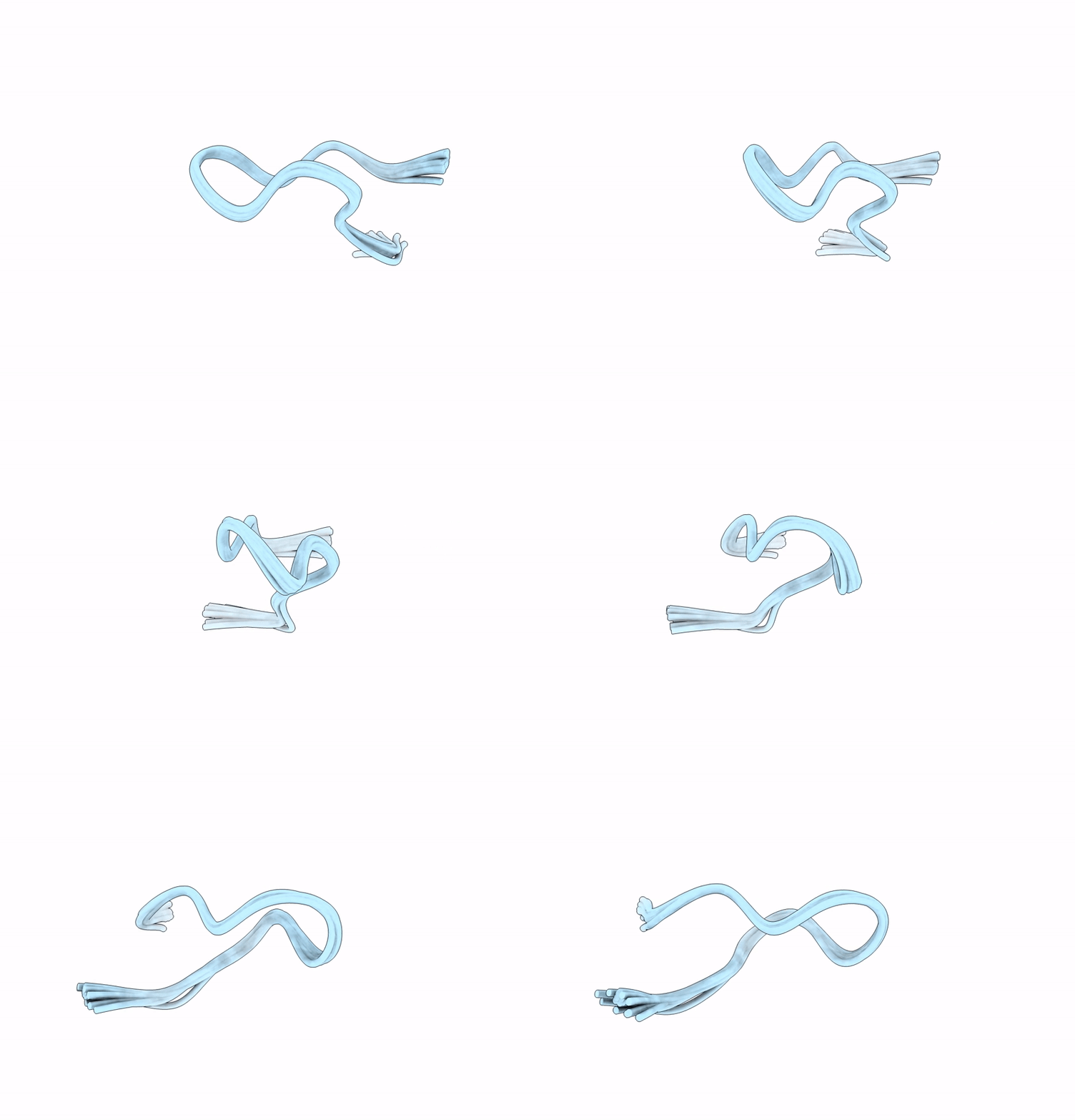 |
Structure Of The Globular Isoform Of The Novel Conotoxin Pnid Derived From Conus Pennaceus
|
 |
Structure Of The Ribbon Isoform Of The Novel Conotoxin Pnid Derived From Conus Pennaceus
|
 |
Solution Nmr Structures Of Dna Minidumbbell Formed With Two Regular Ctttg Pentaloops
|
 |
 |
 |
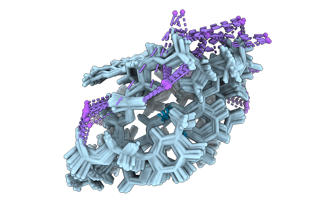
Organism: Homo sapiens
Method: X-RAY DIFFRACTION Resolution:1.11 Å Release Date: 2019-03-20 Classification: CELL CYCLE Ligands: PYZ |
 |
Organism: Homo sapiens
Method: X-RAY DIFFRACTION Resolution:1.29 Å Release Date: 2019-03-20 Classification: CELL CYCLE Ligands: BYZ |
 |
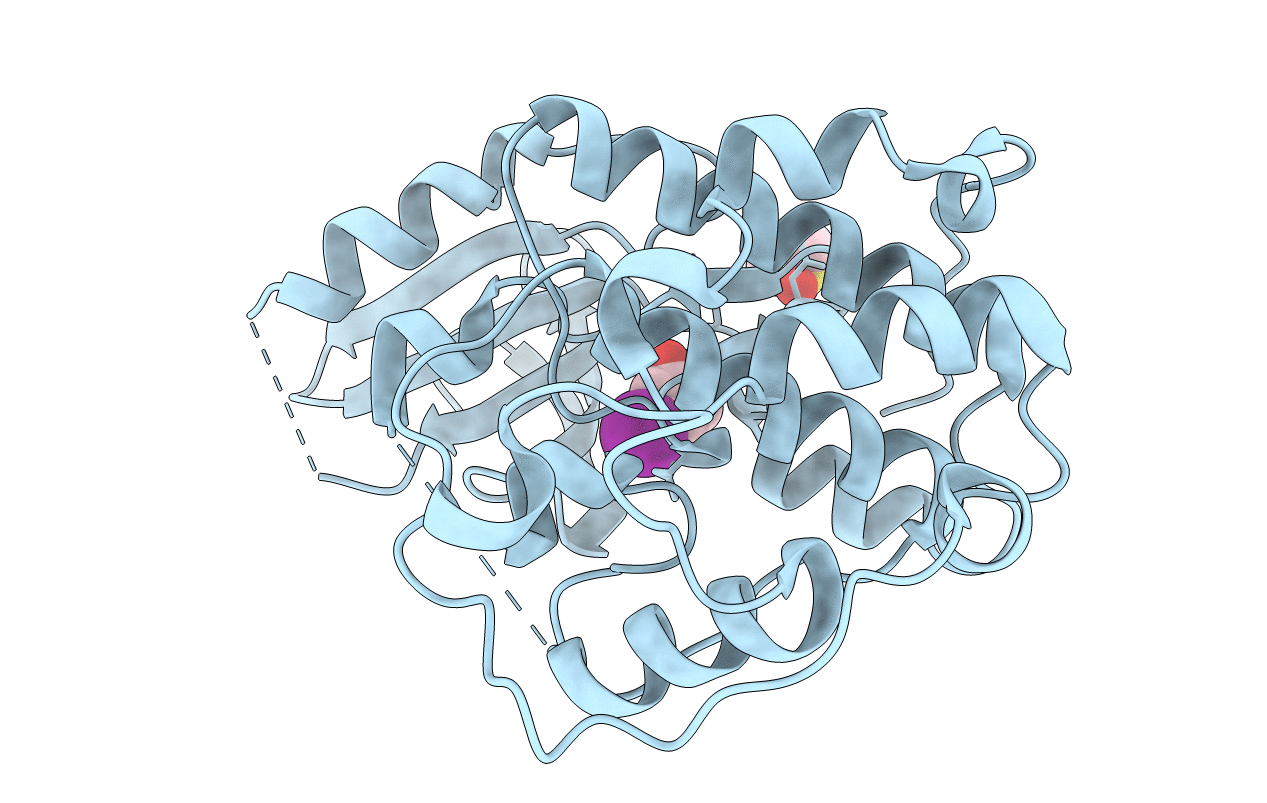
Organism: Homo sapiens
Method: X-RAY DIFFRACTION Resolution:1.18 Å Release Date: 2019-03-20 Classification: CELL CYCLE Ligands: HEW |
 |
Organism: Homo sapiens
Method: X-RAY DIFFRACTION Resolution:1.03 Å Release Date: 2019-03-20 Classification: CELL CYCLE Ligands: DMS, HHQ |
 |
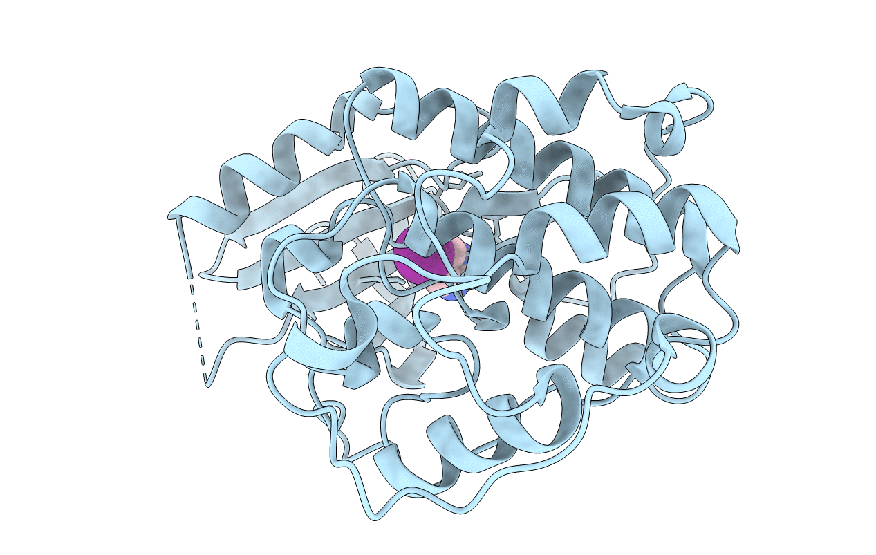
Organism: Homo sapiens
Method: X-RAY DIFFRACTION Resolution:1.00 Å Release Date: 2019-03-20 Classification: CELL CYCLE Ligands: DMS, HGQ |
 |
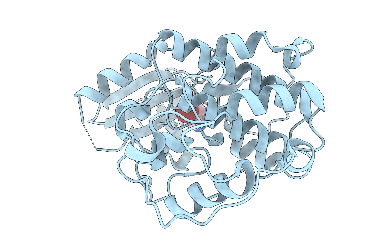
Organism: Homo sapiens
Method: X-RAY DIFFRACTION Resolution:1.13 Å Release Date: 2019-03-20 Classification: CELL CYCLE Ligands: HGW |
 |
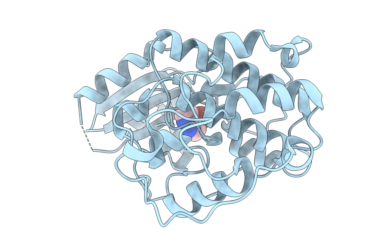
Organism: Homo sapiens
Method: X-RAY DIFFRACTION Resolution:1.12 Å Release Date: 2019-03-20 Classification: CELL CYCLE Ligands: HHN |
 |
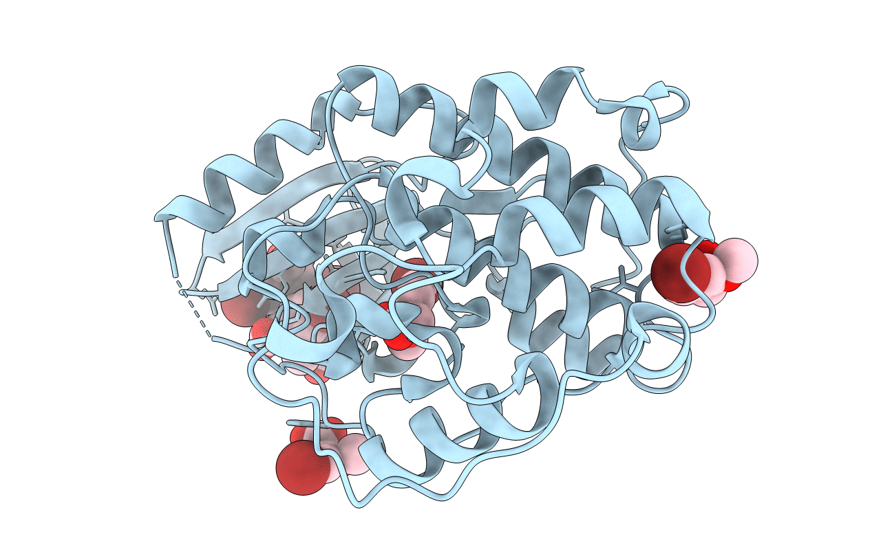
Organism: Homo sapiens
Method: X-RAY DIFFRACTION Resolution:1.73 Å Release Date: 2019-03-20 Classification: CELL CYCLE Ligands: HH8 |
 |
Organism: Homo sapiens
Method: X-RAY DIFFRACTION Resolution:1.07 Å Release Date: 2019-03-20 Classification: CELL CYCLE Ligands: HHT |

