
Search Count: 23
 |
Organism: Trypanosoma brucei
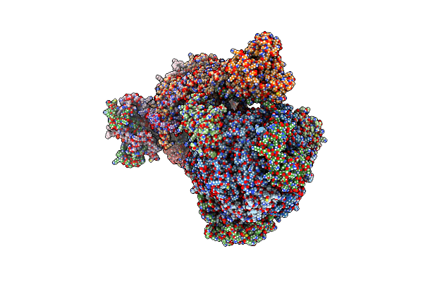
Method: ELECTRON MICROSCOPY Release Date: 2025-03-26 Classification: RIBOSOME Ligands: GTP, MG, SF4, SAM, ACO, ZN, PO4 |
 |
Organism: Trypanosoma brucei brucei
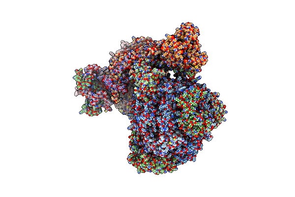
Method: ELECTRON MICROSCOPY Release Date: 2022-12-07 Classification: MEMBRANE PROTEIN Ligands: ATP, MG, CDL, ADP, LMT, Q7G, PEE, PC1, UTP |
 |
Organism: Trypanosoma brucei brucei
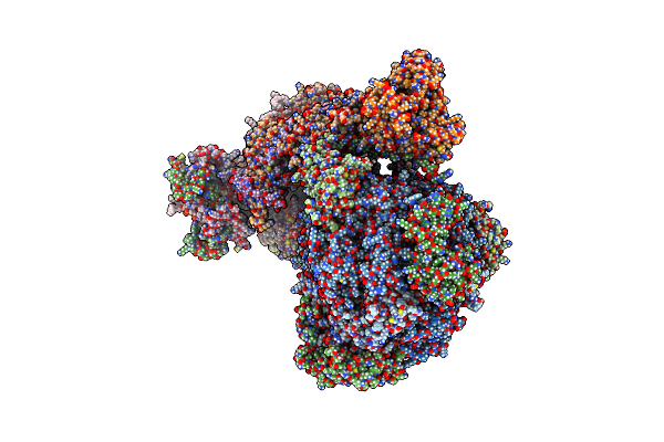
Method: ELECTRON MICROSCOPY Release Date: 2022-10-26 Classification: MEMBRANE PROTEIN Ligands: CDL, LMT, Q7G, PEE, PC1 |
 |
Organism: Trypanosoma brucei brucei
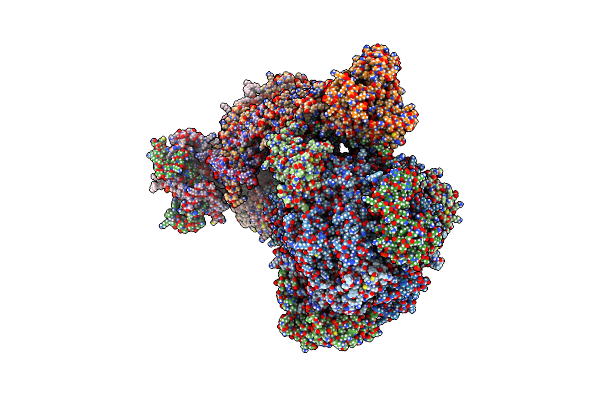
Method: ELECTRON MICROSCOPY Release Date: 2022-10-26 Classification: MEMBRANE PROTEIN |
 |
Organism: Trypanosoma brucei brucei
Method: ELECTRON MICROSCOPY Release Date: 2022-10-26 Classification: MEMBRANE PROTEIN Ligands: UTP |
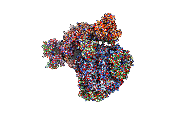 |
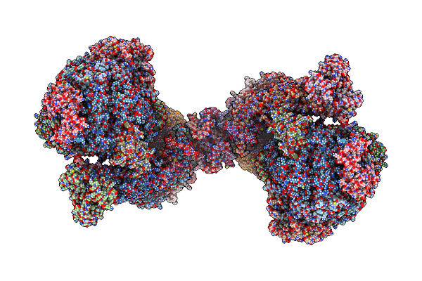
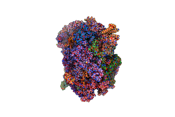
Rotational State 1A Of The Trypanosoma Brucei Mitochondrial Atp Synthase Dimer
Organism: Trypanosoma brucei brucei
Method: ELECTRON MICROSCOPY Release Date: 2022-10-26 Classification: MEMBRANE PROTEIN Ligands: ATP, MG, ADP, UTP, CDL, PEE, LMT, Q7G, PC1 |
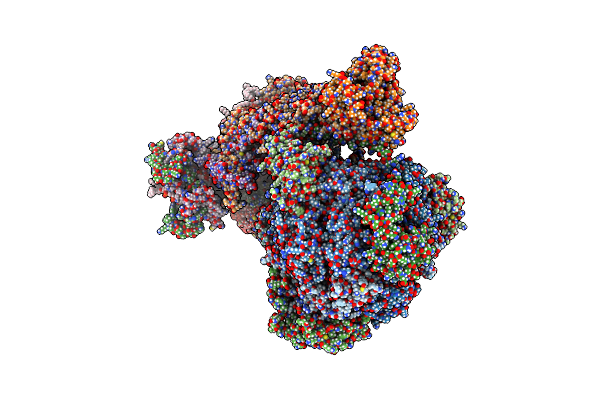 |
Rotational State 1B Of The Trypanosoma Brucei Mitochondrial Atp Synthase Dimer
Organism: Trypanosoma brucei brucei
Method: ELECTRON MICROSCOPY Release Date: 2022-10-26 Classification: MEMBRANE PROTEIN Ligands: ATP, MG, ADP, UTP, CDL, 3PE, PC1, LMT, Q7G |
 |
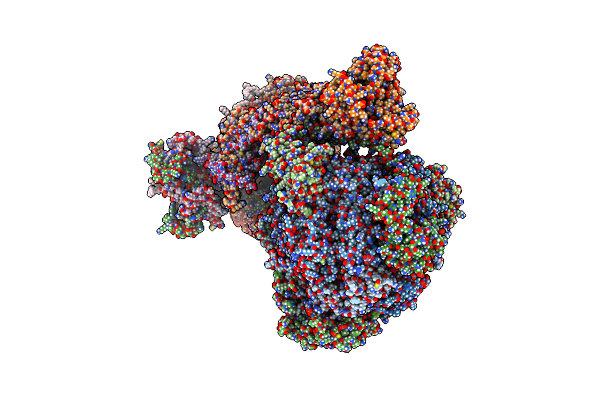
Rotational State 1C Of The Trypanosoma Brucei Mitochondrial Atp Synthase Dimer
Organism: Trypanosoma brucei brucei
Method: ELECTRON MICROSCOPY Release Date: 2022-10-26 Classification: MEMBRANE PROTEIN Ligands: ATP, MG, ADP, UTP, CDL, 3PE, LMT, Q7G, PC1 |
 |
Rotational State 1D Of The Trypanosoma Brucei Mitochondrial Atp Synthase Dimer
Organism: Trypanosoma brucei brucei
Method: ELECTRON MICROSCOPY Release Date: 2022-10-26 Classification: MEMBRANE PROTEIN Ligands: CDL, PEE, PC1, LMT, Q7G, ATP, MG, ADP, UTP |
 |
Rotational State 1E Of The Trypanosoma Brucei Mitochondrial Atp Synthase Dimer
Organism: Trypanosoma brucei brucei
Method: ELECTRON MICROSCOPY Release Date: 2022-10-26 Classification: MEMBRANE PROTEIN Ligands: ATP, MG, ADP, UTP, CDL, PEE, PC1, LMT, Q7G |
 |
Rotational State 2A Of The Trypanosoma Brucei Mitochondrial Atp Synthase Dimer
Organism: Trypanosoma brucei brucei
Method: ELECTRON MICROSCOPY Release Date: 2022-10-26 Classification: MEMBRANE PROTEIN Ligands: CDL, PEE, LMT, Q7G, PC1, UTP, ATP, MG, ADP |
 |
Rotational State 2B Of The Trypanosoma Brucei Mitochondrial Atp Synthase Dimer
Organism: Trypanosoma brucei brucei
Method: ELECTRON MICROSCOPY Release Date: 2022-10-26 Classification: MEMBRANE PROTEIN Ligands: CDL, PEE, LMT, Q7G, PC1, UTP, ATP, MG, ADP |
 |
Rotational State 2C Of The Trypanosoma Brucei Mitochondrial Atp Synthase Dimer
Organism: Trypanosoma brucei brucei
Method: ELECTRON MICROSCOPY Release Date: 2022-10-26 Classification: MEMBRANE PROTEIN Ligands: CDL, PEE, LMT, Q7G, PC1, UTP, ATP, MG, ADP |
 |
Organism: Trypanosoma brucei brucei
Method: ELECTRON MICROSCOPY Release Date: 2022-10-26 Classification: MEMBRANE PROTEIN Ligands: CDL, PEE, LMT, Q7G, PC1, UTP, ATP, MG, ADP |
 |
Rotational State 3 Of The Trypanosoma Brucei Mitochondrial Atp Synthase Dimer
Organism: Trypanosoma brucei brucei
Method: ELECTRON MICROSCOPY Release Date: 2022-10-26 Classification: MEMBRANE PROTEIN Ligands: CDL, 3PE, PC1, LMT, Q7G, ATP, MG, ADP, UTP |
 |
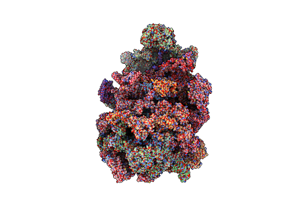
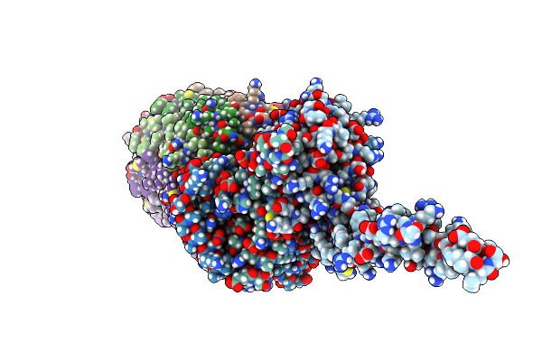
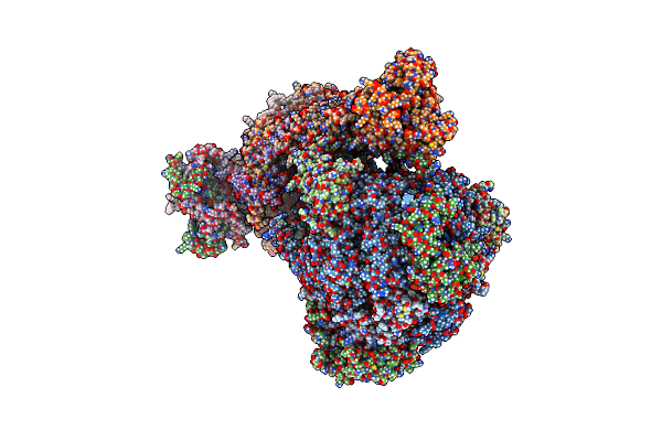
Trypanosoma Brucei Mitochondrial Ribosome Large Subunit Assembly Intermediate
Organism: Trypanosoma brucei
Method: ELECTRON MICROSCOPY Release Date: 2020-12-02 Classification: RIBOSOME Ligands: FES, NAD, ZN, MG, ADP, GTP |
 |
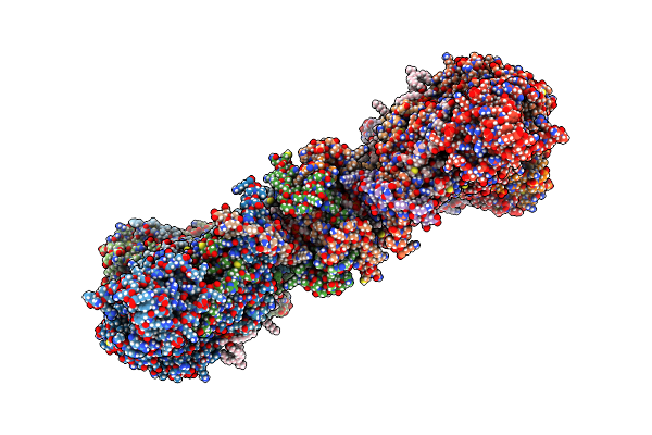
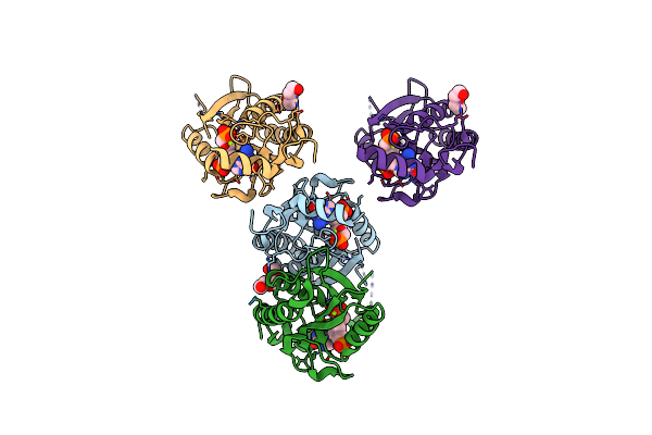
Trypanosoma Brucei Hypoxanthine Guanine Phosphoribosyltransferase In Complex With [(2-((Guanine-9H-Yl)Methyl)Propane-1,3 Diyl)Bis(Oxy)]Bis(Methylene))Diphosphonic Acid
Organism: Trypanosoma brucei brucei
Method: X-RAY DIFFRACTION Resolution:1.76 Å Release Date: 2018-03-14 Classification: TRANSFERASE Ligands: SV2, MG, PEG, TRS, SO4 |
 |
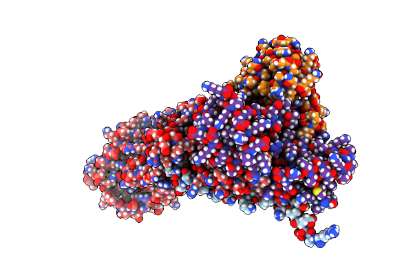
Trypanosoma Brucei Hypoxanthine Guanine Phosphoribosyltransferase In Complex With {[(2S)-3-(2-Amino-6-Oxo-1,6-Dihydro-9H-Purin-9-Yl)Propane-1,2-Diyl]Bis(Oxyethane-2,1-Diyl)}Bis(Phosphonic Acid)
Organism: Trypanosoma brucei brucei
Method: X-RAY DIFFRACTION Resolution:1.80 Å Release Date: 2018-03-14 Classification: TRANSFERASE Ligands: 45T, PEG |
 |
Crystal Structure Of Trypanosoma Brucei Hypoxanthine-Guanine Phosphoribosyltranferase In Complex With (2-{[2-(2-Amino-6-Oxo-1,6-Dihydro-9H-Purin-9-Yl)Ethyl](3-Aminopropyl)Amino}Ethyl)Phosphonic Acid
Organism: Trypanosoma brucei brucei
Method: X-RAY DIFFRACTION Resolution:1.84 Å Release Date: 2018-03-14 Classification: TRANSFERASE Ligands: SO4, MG, PEG, 3L6 |
 |
Trypanosoma Brucei Hypoxanthine Guanine Phosphoribosyltransferase In Complex With [(2-{[2-(2-Amino-6-Oxo-1,6-Dihydro-9H-Purin-9-Yl)Ethyl][(E)-2-Phosphonoethenyl]Amino}Ethoxy)Methyl]Phosphonic Acid
Organism: Trypanosoma brucei brucei
Method: X-RAY DIFFRACTION Resolution:1.99 Å Release Date: 2018-03-14 Classification: TRANSFERASE Ligands: 3L4, MG, PEG |