
Search Count: 63
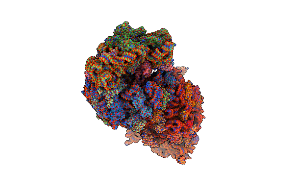 |
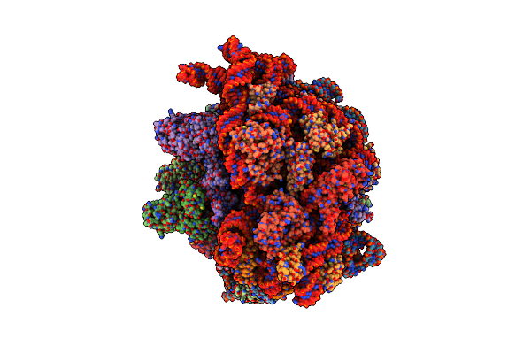
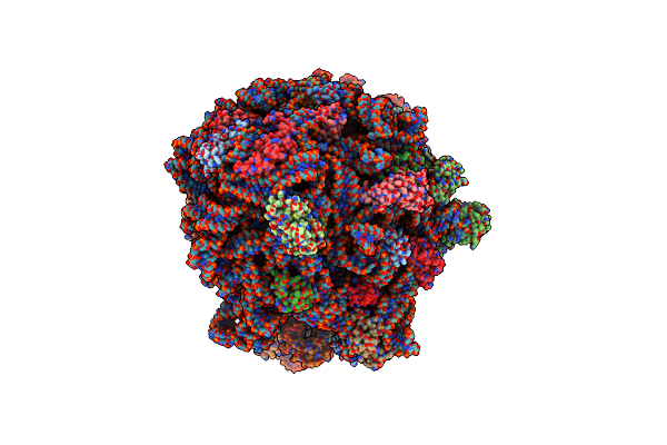
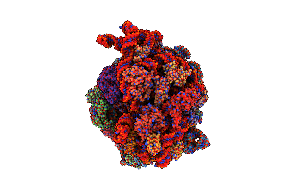
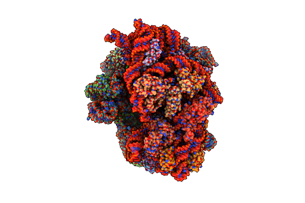
Crystal Structure Of The Wild-Type Thermus Thermophilus 70S Ribosome In Complex With Mrna, A-Site Q230-N5-Methylated Release Factor 1, And P-Site Fmeaaakc-Peptidyl-Trnacys At 2.40A Resolution
Organism: Thermus thermophilus hb8, Escherichia coli, Escherichia phage t4
Method: X-RAY DIFFRACTION Release Date: 2025-06-18 Classification: RIBOSOME Ligands: MG, K, ZN, SF4 |
 |
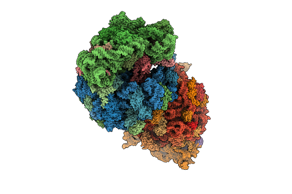
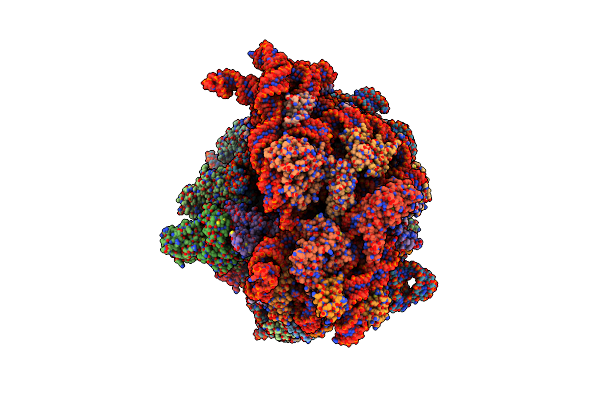
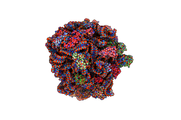
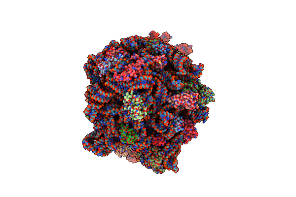
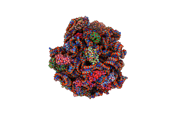
Crystal Structure Of The Wild-Type Thermus Thermophilus 70S Ribosome In Complex With Mrna, A-Site Ggd-Mutant Release Factor 1, And P-Site Fmeaaakc-Peptidyl-Trnacys At 2.55A Resolution
Organism: Thermus thermophilus hb8, Escherichia coli, Escherichia phage t4
Method: X-RAY DIFFRACTION Release Date: 2025-06-18 Classification: RIBOSOME Ligands: MG, K, ZN, SF4 |
 |
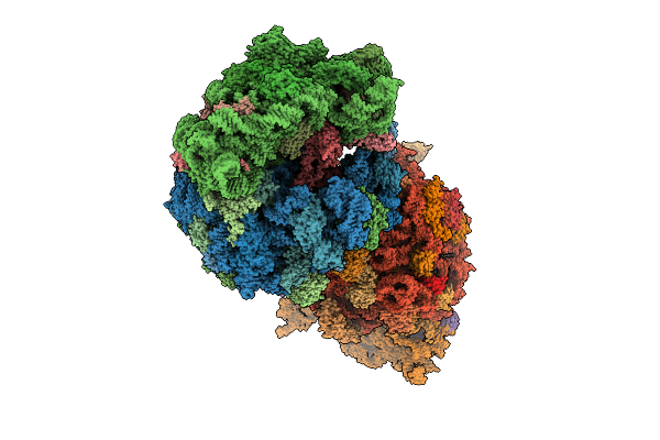
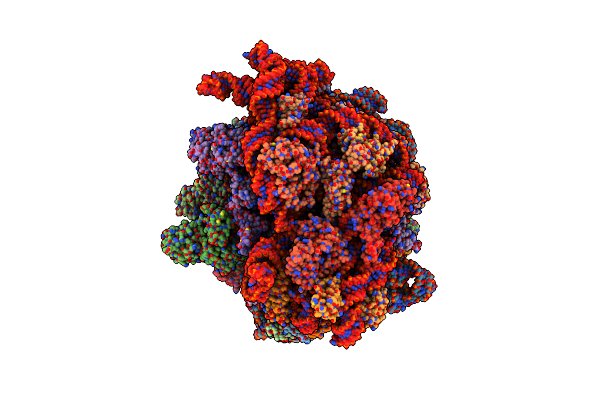
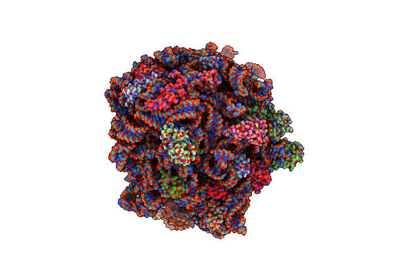
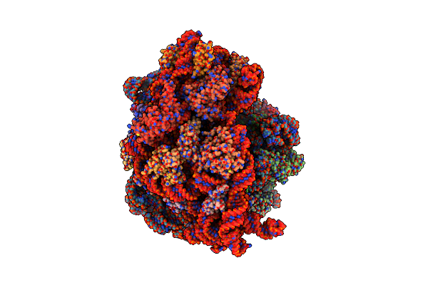
Crystal Structure Of The Wild-Type Thermus Thermophilus 70S Ribosome In Complex With Mrna, A-Site Ggs-Mutant Release Factor 1, And P-Site Fmeaaakc-Peptidyl-Trnacys At 2.80A Resolution
Organism: Thermus thermophilus hb8, Escherichia coli, Escherichia phage t4
Method: X-RAY DIFFRACTION Release Date: 2025-06-18 Classification: RIBOSOME Ligands: MG, K, ZN, SF4 |
 |
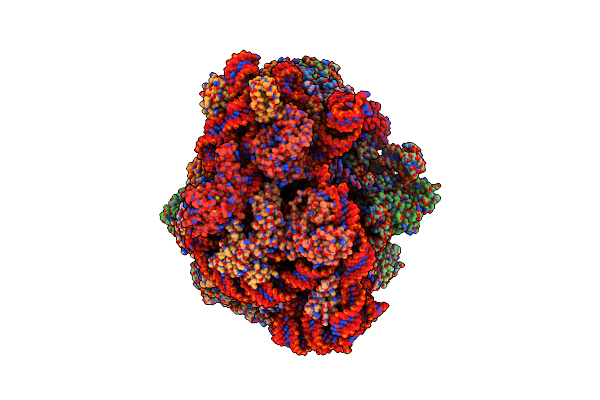
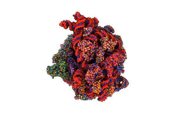
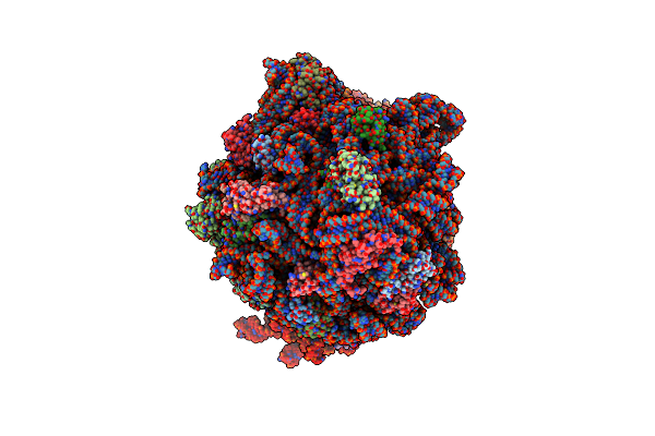
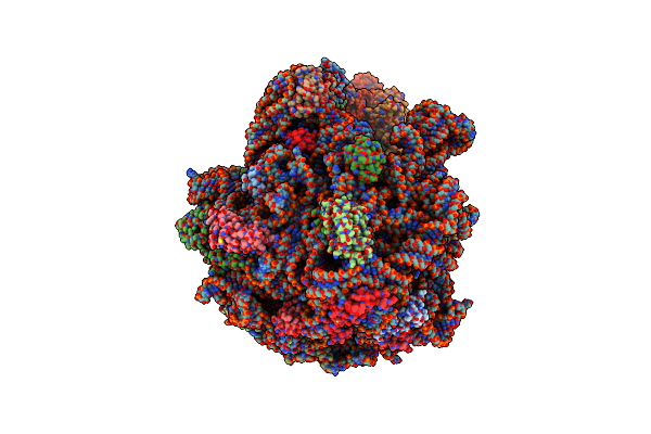
Crystal Structure Of The Wild-Type Thermus Thermophilus 70S Ribosome In Complex With Mrna, A-Site Q230-Unmodified Release Factor 1, And P-Site Fmeaaakc-Peptidyl-Trnacys At 2.70A Resolution
Organism: Thermus thermophilus, Escherichia coli, Escherichia phage t4, Thermus thermophilus hb8
Method: X-RAY DIFFRACTION Release Date: 2025-06-18 Classification: RIBOSOME Ligands: MG, K, ZN, SF4 |
 |
Crystal Structure Of The Wild-Type Thermus Thermophilus 70S Ribosome In Complex With Mrna, A-Site Q230-N5-Methylated Release Factor 1, And P-Site Deacylated-Trnacys At 2.60A Resolution
Organism: Thermus thermophilus hb8, Escherichia coli, Escherichia phage t4
Method: X-RAY DIFFRACTION Release Date: 2025-06-18 Classification: RIBOSOME Ligands: MG, K, ZN, SF4 |
 |
Cryo-Em Structure Of The Wild-Type Thermus Thermophilus 70S Ribosome In Complex With Mrna, A-Site Q230-N5-Methylated Release Factor 1, And P-Site 2'-Deoxy-A76-Fmeaaakc-Peptidyl-Trnacys At 2.13A Resolution
Organism: Thermus thermophilus hb8
Method: ELECTRON MICROSCOPY Release Date: 2025-06-18 Classification: RIBOSOME Ligands: MG, K, ZN, SF4 |
 |
Cryo-Em Structure Of An E. Coli Non-Rotated Ribosome Termination Complex Bound With Aporf3, Rf1, P- And E-Site Trnaphe (Composite State I-B)
Organism: Escherichia coli, Escherichia coli k-12, Escherichia phage t4
Method: ELECTRON MICROSCOPY Release Date: 2024-07-17 Classification: RIBOSOME Ligands: MG, ZN |
 |
Cryo-Em Structure Of An E. Coli Non-Rotated Ribosome Termination Complex Bound With Rf1, P- And E-Site Trnaphe (State I-A)
Organism: Escherichia coli, Escherichia phage t4
Method: ELECTRON MICROSCOPY Release Date: 2024-07-17 Classification: RIBOSOME Ligands: MG, ZN |
 |
Cryo-Em Structure Of An E. Coli Non-Rotated Ribosome Termination Complex Bound With Rf3-Gdpcp, Rf1, P- And E-Site Trnaphe (Composite State Ii-A)
Organism: Escherichia coli, Escherichia coli k-12, Escherichia phage t4
Method: ELECTRON MICROSCOPY Release Date: 2024-07-17 Classification: RIBOSOME Ligands: MG, GCP, ZN |
 |
Cryo-Em Structure Of An E. Coli Non-Rotated Ribosome Termination Complex Bound With Rf1, P- And E-Site Trnaphe (State Ii-D)
Organism: Escherichia coli, Escherichia phage t4
Method: ELECTRON MICROSCOPY Release Date: 2024-07-17 Classification: RIBOSOME Ligands: MG, ZN |
 |
Cryo-Em Structure Of An E. Coli Rotated Ribosome Bound With Rf3-Gdpcp And P/E-Trnaphe (Composite State Ii-B)
Organism: Escherichia coli, Escherichia coli k-12, Escherichia phage t4
Method: ELECTRON MICROSCOPY Release Date: 2024-07-17 Classification: RIBOSOME Ligands: MG, GCP, ZN |
 |
Cryo-Em Structure Of An E. Coli Rotated Ribosome Bound With Rf3-Gdpcp And P/E-Trnaphe (Composite State Ii-C)
Organism: Escherichia coli, Escherichia phage t4
Method: ELECTRON MICROSCOPY Release Date: 2024-07-17 Classification: RIBOSOME Ligands: MG, GCP, ZN |
 |
Cryo-Em Structure Of An E. Coli Rotated Ribosome Complex Bound With Rf3-Ppgpp And P/E-Trnaphe (Composite State I-C)
Organism: Escherichia coli, Escherichia coli k-12, Escherichia phage t4
Method: ELECTRON MICROSCOPY Release Date: 2024-06-26 Classification: RIBOSOME/RNA Ligands: MG, G4P, ZN |
 |
Structure Of The Escherichia Coli 70S Ribosome In Complex With Ef-Tu And Ile-Trnaile(Lau) Bound To The Cognate Aua Codon (Structure I)
Organism: Escherichia coli, Escherichia phage t4
Method: ELECTRON MICROSCOPY Release Date: 2024-03-06 Classification: RIBOSOME Ligands: MG, K, ILE, GCP, ZN |
 |
Structure Of The Escherichia Coli 70S Ribosome In Complex With Ef-Tu And Ile-Trnaile(Lau) Bound To The Near-Cognate Aug Codon (Structure Ii)
Organism: Escherichia coli, Escherichia phage t4
Method: ELECTRON MICROSCOPY Release Date: 2024-03-06 Classification: RIBOSOME Ligands: PAR, MG, ILE, GCP, ZN |
 |
Structure Of The Escherichia Coli 70S Ribosome In Complex With A-Site Trnaile(Lau) Bound To The Cognate Aua Codon (Structure Iii)
Organism: Escherichia coli, Escherichia phage t4
Method: ELECTRON MICROSCOPY Release Date: 2024-03-06 Classification: RIBOSOME Ligands: PAR, MG, K, ILE, ZN |
 |
Structure Of The Escherichia Coli 70S Ribosome In Complex With P-Site Trnaile(Lau) Bound To The Cognate Aua Codon (Structure Iv)
Organism: Escherichia coli, Escherichia phage t4
Method: ELECTRON MICROSCOPY Release Date: 2024-03-06 Classification: RIBOSOME Ligands: MG, ILE, ZN |
 |
Cryo-Em Structure Of The Listeria Innocua 70S Ribosome In Complex With Hpf (Structure I-A)
Organism: Listeria innocua
Method: ELECTRON MICROSCOPY Release Date: 2024-02-28 Classification: RIBOSOME Ligands: MG, ZN |
 |
Cryo-Em Structure Of The Listeria Innocua 70S Ribosome (Head-Swiveled) In Complex With Pe/E-Trna (Structure I-B)
Organism: Escherichia coli, Escherichia phage t4, Listeria innocua
Method: ELECTRON MICROSCOPY Release Date: 2024-02-28 Classification: RIBOSOME Ligands: MG, ZN |
 |
Cryo-Em Structure Of The Ratcheted Listeria Innocua 70S Ribosome In Complex With P/E-Trna (Structure Ii-A)
Organism: Escherichia phage t4, Listeria innocua, Escherichia coli
Method: ELECTRON MICROSCOPY Release Date: 2024-02-28 Classification: RIBOSOME Ligands: MG, ZN, K |