
Search Count: 151
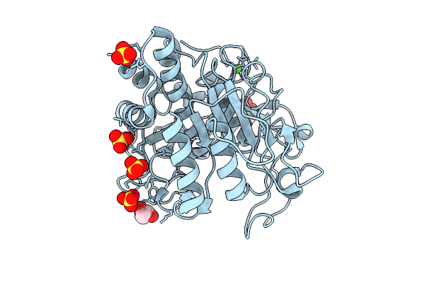 |
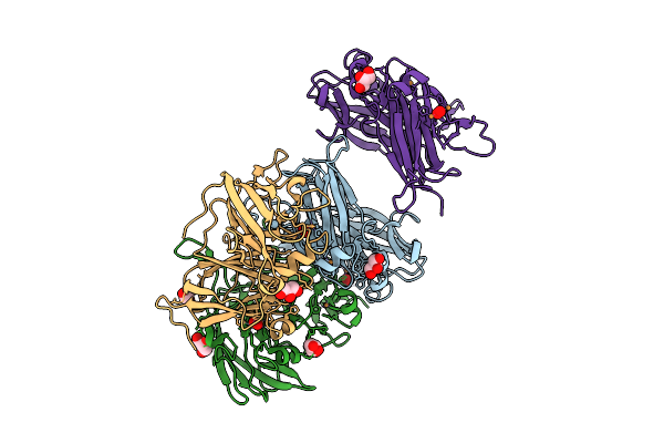
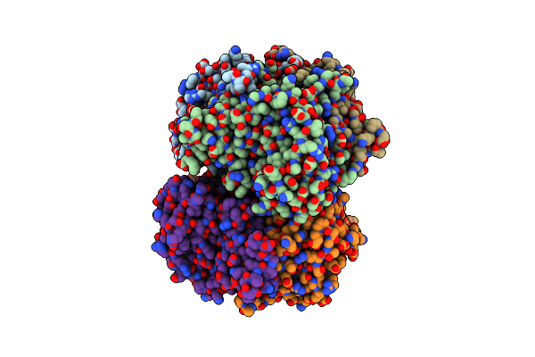
Stmpr1, Stenotrophomonas Maltophilia Protease 1, 36 Kda Alkine Serine Protease
Organism: Stenotrophomonas maltophilia
Method: X-RAY DIFFRACTION Release Date: 2025-08-06 Classification: HYDROLASE Ligands: GOL, CA, SO4 |
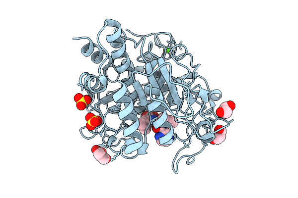 |
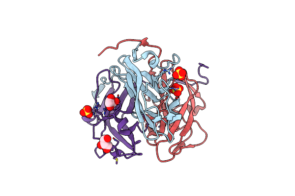
Stmpr1, Stenotrophomonas Maltophilia Protease 1, 36 Kda Alkine Serine Protease In Complex With Bortezomib
Organism: Stenotrophomonas maltophilia
Method: X-RAY DIFFRACTION Release Date: 2025-08-06 Classification: HYDROLASE Ligands: BO2, GOL, SO4, CA |
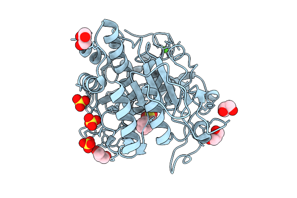 |
Stmpr1, Stenotrophomonas Maltophilia Protease 1, 36 Kda Alkine Serine Protease In Complex With Pmsf
Organism: Stenotrophomonas maltophilia
Method: X-RAY DIFFRACTION Release Date: 2025-08-06 Classification: HYDROLASE Ligands: GOL, PMS, SO4, CA |
 |
Stmpr1, Stenotrophomonas Maltophilia Protease 1, 36 Kda Alkine Serine Protease In Complex With Chymostatin
Organism: Stenotrophomonas maltophilia
Method: X-RAY DIFFRACTION Release Date: 2025-08-06 Classification: HYDROLASE Ligands: CA, SO4, GOL, A1I1B |
 |
Stmpr1, Stenotrophomonas Maltophilia Protease 1, 36 Kda Alkine Serine Protease In Complex With Leupeptin
Organism: Stenotrophomonas maltophilia, Synthetic construct
Method: X-RAY DIFFRACTION Release Date: 2025-08-06 Classification: HYDROLASE Ligands: CA, SO4, GOL |
 |
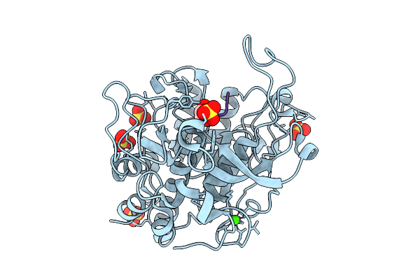
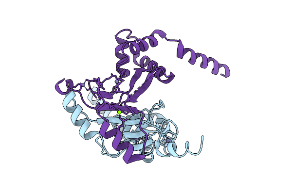
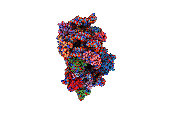
Mutant H286T Crystal Structure Of Two-Domain Bacterial Laccase From The Actinobacterium Streptomyces Carpinensis Vkm Ac-1300
Organism: Streptomyces carpinensis
Method: X-RAY DIFFRACTION Release Date: 2025-08-06 Classification: OXIDOREDUCTASE Ligands: CU, OXY, GOL |
 |
Organism: Bacillus cereus vd154
Method: X-RAY DIFFRACTION Release Date: 2025-07-23 Classification: METAL BINDING PROTEIN Ligands: ZN, MG |
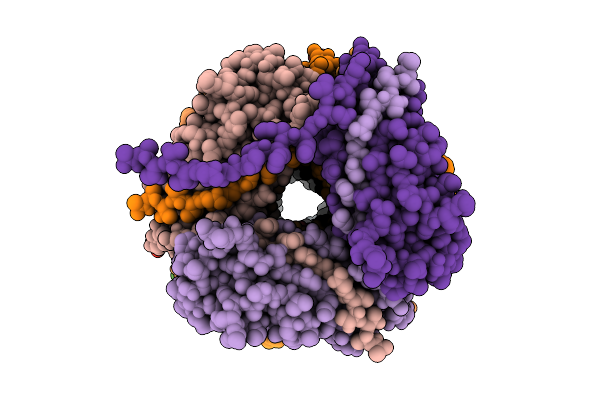 |
Cryo-Em Structure Of The Type I Pilus From Escherichia Coli And The Surrounding Water Network
Organism: Escherichia coli (strain k12)
Method: ELECTRON MICROSCOPY Release Date: 2025-07-16 Classification: CELL ADHESION |
 |
Organism: Beggiatoa leptomitoformis
Method: X-RAY DIFFRACTION Resolution:2.33 Å Release Date: 2025-03-05 Classification: OXIDOREDUCTASE Ligands: FE |
 |
Organism: Escherichia coli o157:h7, Branchiostoma lanceolatum
Method: X-RAY DIFFRACTION Resolution:2.65 Å Release Date: 2024-12-25 Classification: FLUORESCENT PROTEIN Ligands: ILE, GOL, CL |
 |
Organism: Escherichia coli, Escherichia coli o157:h7
Method: X-RAY DIFFRACTION Resolution:1.62 Å Release Date: 2024-12-25 Classification: FLUORESCENT PROTEIN Ligands: ILE, GOL, CL |
 |
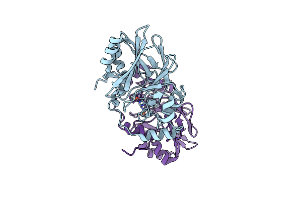
Crystal Structure Of Two-Domain Laccase Mutant M199A/D268N From Streptomyces Griseoflavus
Organism: Streptomyces griseoflavus
Method: X-RAY DIFFRACTION Resolution:2.00 Å Release Date: 2024-04-10 Classification: OXIDOREDUCTASE Ligands: CU, OXY, CA |
 |
Crystal Structure Of Two-Domain Laccase Mutant M199G/R240H/D268N From Streptomyces Griseoflavus
Organism: Streptomyces griseoflavus
Method: X-RAY DIFFRACTION Resolution:2.20 Å Release Date: 2024-04-10 Classification: OXIDOREDUCTASE Ligands: OXY, CU, PER |
 |
Organism: Escherichia phage t5
Method: X-RAY DIFFRACTION Resolution:2.00 Å Release Date: 2024-01-17 Classification: HYDROLASE Ligands: GOL |
 |
Organism: Escherichia phage t5
Method: X-RAY DIFFRACTION Resolution:2.10 Å Release Date: 2024-01-17 Classification: HYDROLASE Ligands: SO4, CL, GOL |
 |
Organism: Staphylococcus aureus subsp. aureus nctc 8325
Method: ELECTRON MICROSCOPY Release Date: 2023-12-27 Classification: RIBOSOME |
 |
Organism: Cytaeis uchidae
Method: X-RAY DIFFRACTION Resolution:1.45 Å Release Date: 2023-12-27 Classification: FLUORESCENT PROTEIN Ligands: CL |
 |
Organism: Cytaeis uchidae
Method: X-RAY DIFFRACTION Resolution:1.80 Å Release Date: 2023-12-27 Classification: FLUORESCENT PROTEIN Ligands: CL |
 |
Organism: Cytaeis uchidae
Method: X-RAY DIFFRACTION Resolution:1.60 Å Release Date: 2023-12-27 Classification: FLUORESCENT PROTEIN Ligands: SO4, CL |
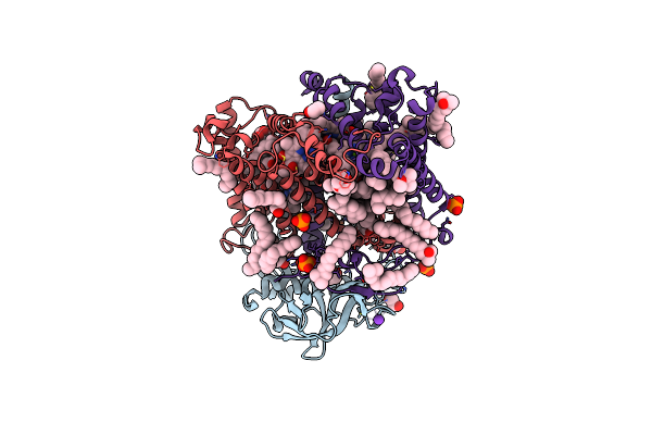 |
Double Mutant A(L37)C/S(L99)C Structure Of Photosynthetic Reaction Center From Cereibacter Sphaeroides Strain Rv
Organism: Cereibacter sphaeroides 2.4.1
Method: X-RAY DIFFRACTION Resolution:2.60 Å Release Date: 2023-11-22 Classification: PHOTOSYNTHESIS Ligands: LDA, UNL, PO4, EDO, K, BCL, BPH, U10, DIO, HTO, CDL, FE, SPN |