
Search Count: 17
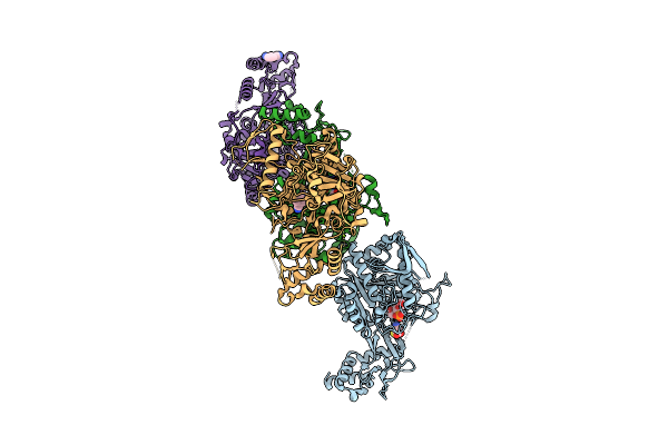 |
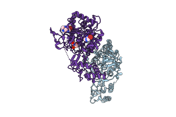
The Structure Of Aspergillus Fumigatus Udp-Glcnac Pyrophosphorylase In Complex With A Fragment
Organism: Aspergillus fumigatus
Method: X-RAY DIFFRACTION Release Date: 2025-07-30 Classification: TRANSFERASE Ligands: GN1, A1IIG, 71G, MG, CL |
 |
The Structure Of Aspergillus Fumigatus Udp-Glcnac Pyrophosphorylase In Complex With A Fragment
Organism: Aspergillus fumigatus
Method: X-RAY DIFFRACTION Release Date: 2025-07-30 Classification: TRANSFERASE Ligands: A1IHU, GN1 |
 |
The Structure Of Aspergillus Fumigatus Udp-Glcnac Pyrophosphorylase In Complex With A Fragment
Organism: Aspergillus fumigatus
Method: X-RAY DIFFRACTION Release Date: 2025-07-30 Classification: TRANSFERASE Ligands: GN1, YRL, MG |
 |
The Structure Of Aspergillus Fumigatus Udp-Glcnac Pyrophosphorylase In Complex With A Fragment
Organism: Aspergillus fumigatus
Method: X-RAY DIFFRACTION Release Date: 2025-07-30 Classification: TRANSFERASE Ligands: YRL, GN1, PO4 |
 |
The Structure Of Aspergillus Fumigatus Udp-Glcnac Pyrophosphorylase In Complex With A Fragment
Organism: Aspergillus fumigatus
Method: X-RAY DIFFRACTION Release Date: 2025-07-30 Classification: TRANSFERASE Ligands: A1IIQ, GN1, A1IIG |
 |
The Structure Of Aspergillus Fumigatus Udp-Glcnac Pyrophosphorylase In Complex With Fragments
Organism: Aspergillus fumigatus
Method: X-RAY DIFFRACTION Release Date: 2025-07-30 Classification: TRANSFERASE Ligands: GN1, A1IHU, A1IIR, CL |
 |
Crystal Structure Of Aspergillus Fumigatus Udp-N-Acetylglucosamine Pyrophosphorylase In Complex With Glcnac-1P
Organism: Aspergillus fumigatus af293
Method: X-RAY DIFFRACTION Resolution:2.28 Å Release Date: 2020-04-01 Classification: TRANSFERASE Ligands: PO4, GN1 |
 |
Structure Of Conglutinin Carbohydrate Recognition Domain With Glcnac-Alpha-1-Phosphate Bound
Organism: Bos taurus
Method: X-RAY DIFFRACTION Resolution:1.00 Å Release Date: 2019-10-09 Classification: IMMUNE SYSTEM Ligands: CA, GN1 |
 |
Structure Of The Lactococcal Phage 1358 Receptor Binding Protein In Complex With Glcnac-1P
Organism: Lactococcus phage 1358
Method: X-RAY DIFFRACTION Resolution:2.15 Å Release Date: 2014-10-15 Classification: VIRAL PROTEIN Ligands: GN1 |
 |
Organism: Bifidobacterium longum
Method: X-RAY DIFFRACTION Resolution:2.16 Å Release Date: 2014-05-14 Classification: TRANSFERASE Ligands: GN1 |
 |
N-Acetylhexosamine 1-Phosphate Kinase In Complex With Glcnac-1-Phosphate And Adp
Organism: Bifidobacterium longum
Method: X-RAY DIFFRACTION Resolution:1.94 Å Release Date: 2014-05-14 Classification: TRANSFERASE Ligands: GN1, ADP, MG |
 |
Organism: Mycobacterium tuberculosis
Method: X-RAY DIFFRACTION Resolution:1.98 Å Release Date: 2014-04-16 Classification: TRANSFERASE Ligands: ATP, MG, GN1, CO |
 |
Crystal Structure Of Glmu From Mycobacterium Tuberculosis In Complex With Uridine-Diphosphate-N-Acetylglucosamine And Pyrophosphate Snapshot 2
Organism: Mycobacterium tuberculosis
Method: X-RAY DIFFRACTION Resolution:2.04 Å Release Date: 2013-08-07 Classification: TRANSFERASE Ligands: UD1, GN1, POP, MG, CO |
 |
Crystal Structure Of Glmu From Mycobacterium Tuberculosis In Complex With Glucosamine-1-Phosphate
Organism: Mycobacterium tuberculosis
Method: X-RAY DIFFRACTION Resolution:2.60 Å Release Date: 2013-03-13 Classification: TRANSFERASE Ligands: GN1, CO, MG |
 |
N-Acetyl Glucosamine 1-Phosphate Uridyltransferase From Mycobacterium Tuberculosis Complex With N-Acetyl Glucosamine 1-Phosphate
Organism: Mycobacterium tuberculosis
Method: X-RAY DIFFRACTION Resolution:2.75 Å Release Date: 2008-07-15 Classification: TRANSFERASE Ligands: GN1 |
 |
Organism: Escherichia coli
Method: X-RAY DIFFRACTION Resolution:2.54 Å Release Date: 2007-06-19 Classification: TRANSFERASE Ligands: GN1, CO, DCA, UD1, MG, SO4 |
 |
Crystal Structure Of Uridine-Diphospho-N-Acetylglucosamine Pyrophosphorylase From Candida Albicans, In The Substrate-Binding Form
Organism: Candida albicans
Method: X-RAY DIFFRACTION Resolution:2.30 Å Release Date: 2007-05-22 Classification: TRANSFERASE Ligands: GN1, SO4, MG, GOL |