
Search Count: 89
 |
Organism: Agrobacterium fabrum (strain c58 / atcc 33970)
Method: X-RAY DIFFRACTION Resolution:1.65 Å Release Date: 2016-03-23 Classification: LYASE Ligands: GOL, FMT |
 |
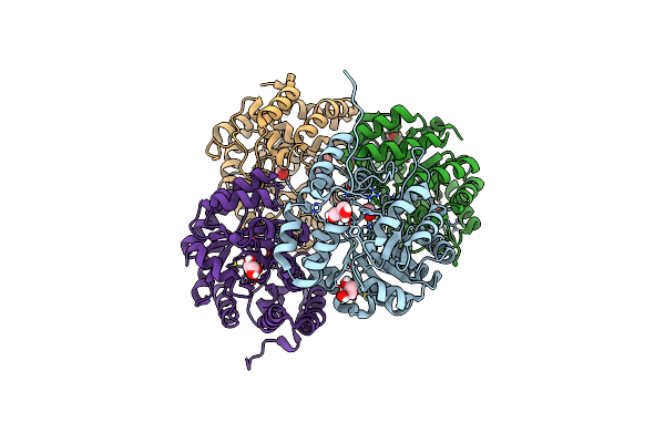
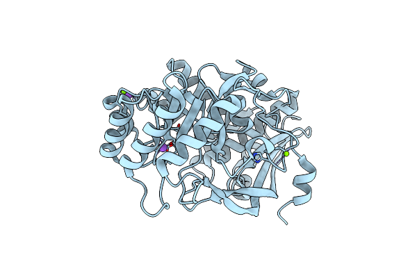
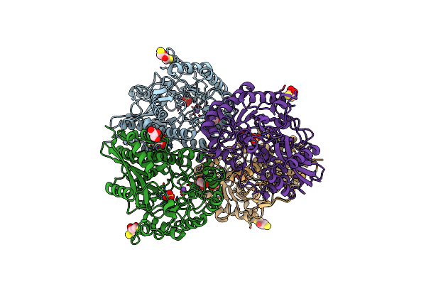
Crystal Structure Of Keto-Deoxy-D-Galactarate Dehydratase Complexed With 2-Oxoadipic Acid
Organism: Agrobacterium fabrum (strain c58 / atcc 33970)
Method: X-RAY DIFFRACTION Resolution:2.10 Å Release Date: 2016-03-23 Classification: LYASE Ligands: OOG, FMT |
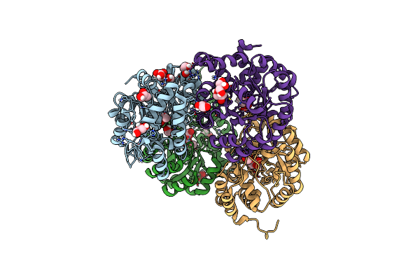 |
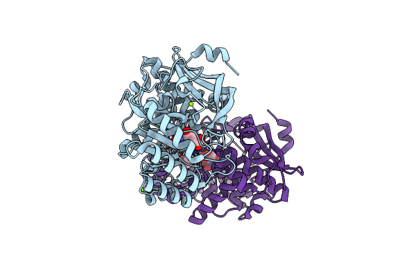
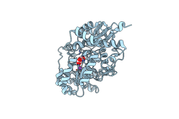
Crystal Structure Of Keto-Deoxy-D-Galactarate Dehydratase Complexed With Pyruvate
Organism: Agrobacterium fabrum (strain c58 / atcc 33970)
Method: X-RAY DIFFRACTION Resolution:1.50 Å Release Date: 2016-03-23 Classification: LYASE Ligands: PYR, GOL, FMT |
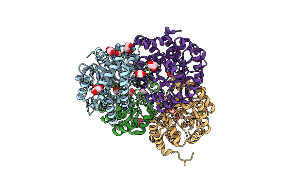 |
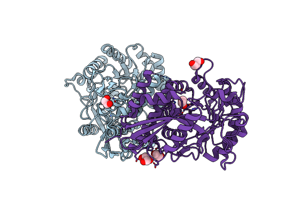
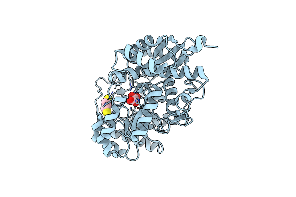
Crystal Structure Of Keto-Deoxy-D-Galactarate Dehydratase Complexed With Pyruvate
Organism: Agrobacterium tumefaciens
Method: X-RAY DIFFRACTION Resolution:1.50 Å Release Date: 2014-12-17 Classification: LYASE Ligands: GOL, FMT |
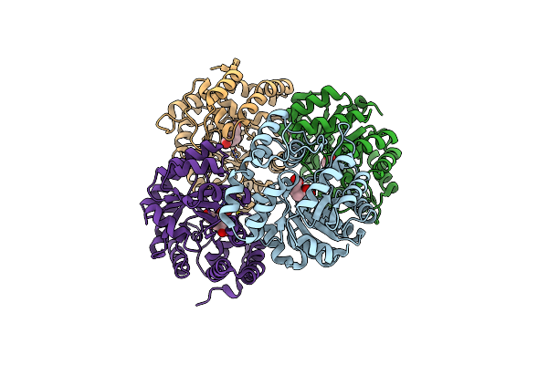 |
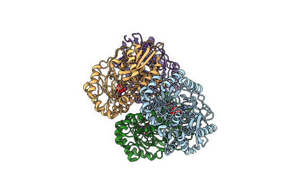
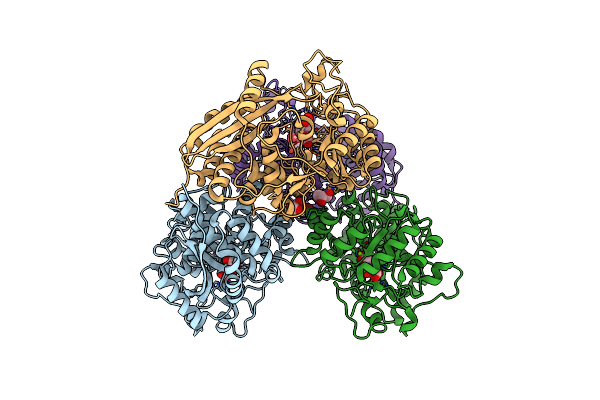
Crystal Structure Of Keto-Deoxy-D-Galactarate Dehydratase Complexed With 2-Oxoadipic Acid
Organism: Agrobacterium tumefaciens
Method: X-RAY DIFFRACTION Resolution:2.10 Å Release Date: 2014-12-17 Classification: LYASE Ligands: OOG, FMT |
 |
Crystal Structure Of D-Glucarate Dehydratase From Agrobacterium Tumefaciens Complexed With Magnesium
Organism: Agrobacterium tumefaciens
Method: X-RAY DIFFRACTION Resolution:1.70 Å Release Date: 2013-10-30 Classification: ISOMERASE Ligands: CL, MG, NA |
 |
Crystal Structure Of D-Glucarate Dehydratase From Agrobacterium Tumefaciens Complexed With Magnesium, L-Xylarohydroxamate And L-Lyxarohydroxamate
Organism: Agrobacterium fabrum
Method: X-RAY DIFFRACTION Resolution:1.65 Å Release Date: 2013-09-18 Classification: ISOMERASE Ligands: MG, XYH, LLH, CL, PGE |
 |
Structure Of Putative Glucarate Dehydratase From Acidaminococcus Sp. D21 With Unusual Static Disorder
Organism: Acidaminococcus sp. d21
Method: X-RAY DIFFRACTION Resolution:1.84 Å Release Date: 2013-02-13 Classification: LYASE Ligands: EDO, CL, GOL |
 |
Crystal Structure Of Enolase Pfl01_3283 (Target Efi-502286) From Pseudomonas Fluorescens Pf0-1 With Bound Magnesium, Potassium And Tartrate
Organism: Pseudomonas fluorescens
Method: X-RAY DIFFRACTION Resolution:2.20 Å Release Date: 2013-01-30 Classification: ISOMERASE Ligands: MG, TLA, K, BCT |
 |
Crystal Structure Of Glucdrp From E. Coli K-12 Mg1655 (Efi Target Efi-506058)
Organism: Escherichia coli
Method: X-RAY DIFFRACTION Resolution:2.80 Å Release Date: 2013-01-16 Classification: LYASE Ligands: CIT, GOL |
 |
Crystal Structure Of A Putative D-Glucarate Dehydratase From Pseudomonas Mendocina Ymp
Organism: Pseudomonas mendocina
Method: X-RAY DIFFRACTION Resolution:2.20 Å Release Date: 2012-11-07 Classification: LYASE Ligands: GOL |
 |
Crystal Structure Of D-Glucarate Dehydratase From Agrobacterium Tumefaciens Complexed With Magnesium And L-Tartrate
Organism: Agrobacterium tumefaciens
Method: X-RAY DIFFRACTION Resolution:1.70 Å Release Date: 2012-10-24 Classification: ISOMERASE Ligands: TLA, GOL, MG, NA, PGE, EDO |
 |
Crystal Structure Of D-Glucarate Dehydratase From Agrobacterium Tumefaciens Complexed With Magnesium And L-Lyxarohydroxamate
Organism: Agrobacterium tumefaciens
Method: X-RAY DIFFRACTION Resolution:1.80 Å Release Date: 2012-10-24 Classification: ISOMERASE Ligands: MG, LLH, PG4 |
 |
Crystal Structure Of The Heterotetrameric Complex Of Glucd And Glucdrp From E. Coli K-12 Mg1655 (Efi Target Efi-506058)
Organism: Escherichia coli
Method: X-RAY DIFFRACTION Resolution:2.10 Å Release Date: 2012-09-12 Classification: LYASE/LYASE Ligands: MG, SO4, GOL, PEG, CIT, P6G |
 |
Crystal Structure Of A Glucarate Dehydratase Related Protein, From Actinobacillus Succinogenes, Target Efi-502312, With Sodium And Sulfate Bound, Ordered Loop
Organism: Actinobacillus succinogenes
Method: X-RAY DIFFRACTION Resolution:1.70 Å Release Date: 2012-08-15 Classification: LYASE Ligands: SO4, NA, GOL, DTU, DTT |
 |
Crystal Structure Of Enolase Msmeg_6132 (Target Efi-502282) From Mycobacterium Smegmatis Str. Mc2 155 Complexed With Formate And Acetate
Organism: Mycobacterium smegmatis str. mc2 155
Method: X-RAY DIFFRACTION Resolution:1.90 Å Release Date: 2012-01-25 Classification: LYASE Ligands: FMT, NA, CL, ACT |
 |
Crystal Structure Of Enolase Msmeg_6132 (Target Efi-502282) From Mycobacterium Smegmatis Str. Mc2 155 Complexed With Tartrate
Organism: Mycobacterium smegmatis str. mc2 155
Method: X-RAY DIFFRACTION Resolution:2.00 Å Release Date: 2012-01-25 Classification: LYASE Ligands: TLA, DTT, CL |
 |
Crystal Structure Of The Mutant S29G.P34A Of D-Glucarate Dehydratase From Escherichia Coli Complexed With Product 5-Keto-4-Deoxy-D-Glucarate
Organism: Escherichia coli
Method: X-RAY DIFFRACTION Resolution:2.00 Å Release Date: 2011-12-28 Classification: LYASE Ligands: MG, GLR, GOL |
 |
Crystal Structure Of The Mutant P34A Of D-Glucarate Dehydratase From Escherichia Coli Complexed With Product 5-Keto-4-Deoxy-D-Glucarate
Organism: Escherichia coli
Method: X-RAY DIFFRACTION Resolution:2.23 Å Release Date: 2011-12-28 Classification: LYASE Ligands: MG, GLR, GOL |
 |
Crystal Structure Of D-Glucarate Dehydratase Related Protein From Actinobacillus Succinogenes Complexed With D-Glucarate
Organism: Actinobacillus succinogenes
Method: X-RAY DIFFRACTION Resolution:1.90 Å Release Date: 2011-11-02 Classification: ISOMERASE Ligands: MG, GKR |