
Search Count: 27
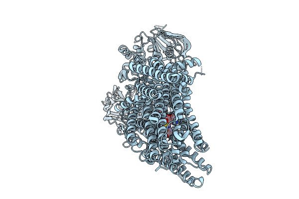 |
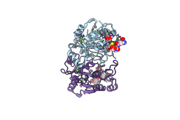
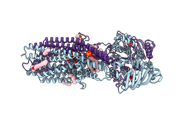
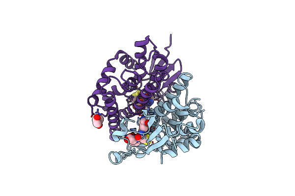
Organism: Bos taurus
Method: ELECTRON MICROSCOPY Release Date: 2025-10-01 Classification: PROTEIN TRANSPORT Ligands: GDS |
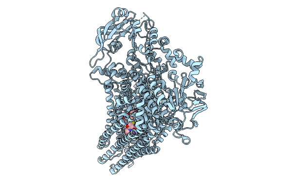 |
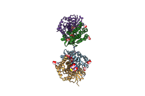
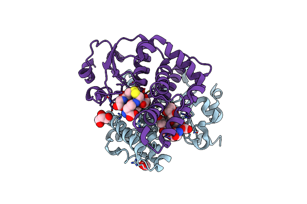
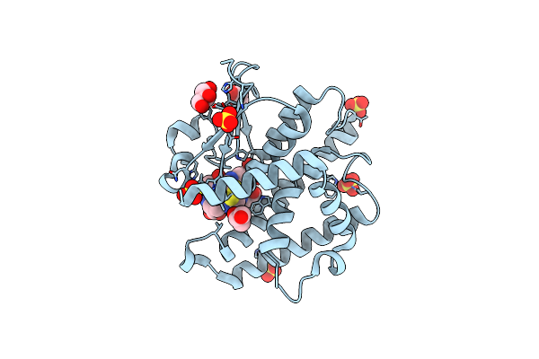
Organism: Saccharomyces cerevisiae
Method: ELECTRON MICROSCOPY Release Date: 2025-09-24 Classification: MEMBRANE PROTEIN Ligands: GDS |
 |
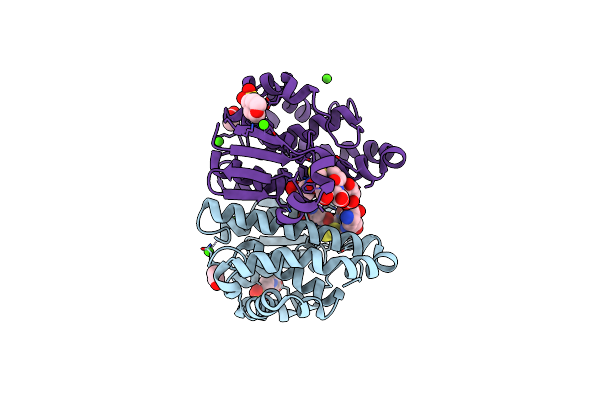
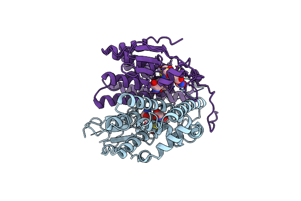
Organism: Arabidopsis thaliana
Method: ELECTRON MICROSCOPY Release Date: 2022-04-13 Classification: MEMBRANE PROTEIN Ligands: GDS |
 |
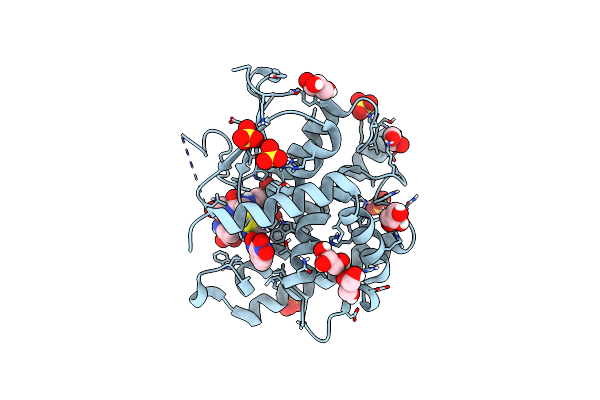
Glutathione-S-Transferase Glig Mutant K127G In Complex With Oxidized Glutathione
Organism: Aspergillus fumigatus a1163
Method: X-RAY DIFFRACTION Resolution:1.50 Å Release Date: 2021-05-12 Classification: BIOSYNTHETIC PROTEIN Ligands: GDS, EDO |
 |
Acylintermediate Of Glutathione And The Mature Primitive Phytochelatin Synthase Alr0975 From Nostoc Pcc 7120 At Atomic Resolution.
Organism: Nostoc sp. pcc 7120
Method: X-RAY DIFFRACTION Resolution:1.09 Å Release Date: 2020-12-02 Classification: TRANSFERASE Ligands: GDS, CA |
 |
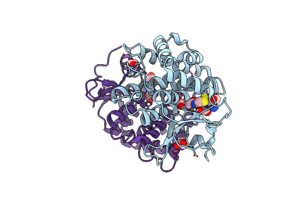
Crystal Structure Of A Nu-Class Glutathione-S-Transferase From Pseudomonas Aeruginosa Pacs2 Bound To Glutathione
Organism: Pseudomonas aeruginosa
Method: X-RAY DIFFRACTION Resolution:1.30 Å Release Date: 2020-09-16 Classification: TRANSFERASE Ligands: GDS, EDO, GOL |
 |
Organism: Homo sapiens
Method: X-RAY DIFFRACTION Resolution:1.55 Å Release Date: 2018-08-22 Classification: TRANSFERASE Ligands: GSH, GDS, MES, ACT, CA |
 |
Ure2P5 From Phanerochaete Chrysosporium Cocrystallized With 1-(S-Glutathionyl)-2,4-Dinitrobenzene.
Organism: Phanerochaete chrysosporium
Method: X-RAY DIFFRACTION Resolution:2.03 Å Release Date: 2017-05-24 Classification: TRANSFERASE Ligands: GDS, NIN, SO4, GOL |
 |
Organism: Moorea producens 3l
Method: X-RAY DIFFRACTION Resolution:2.09 Å Release Date: 2016-10-19 Classification: TRANSFERASE,LYASE Ligands: SAH, GDS |
 |
Crystal Structure Of Glutathione Transferase Ure2P4 From Phanerochaete Chrysosporium In Complex With Oxidized Glutathione.
Organism: Phanerochaete chrysosporium
Method: X-RAY DIFFRACTION Resolution:1.80 Å Release Date: 2015-09-30 Classification: TRANSFERASE Ligands: GDS, NA |
 |
Crystal Structure Of The Glutathione Transferase Ure2P6 From Phanerochaete Chrysosporium In Complex With Oxidized Glutathione.
Organism: Phanerochaete chrysosporium
Method: X-RAY DIFFRACTION Resolution:2.00 Å Release Date: 2015-09-30 Classification: TRANSFERASE Ligands: GDS |
 |
Crystal Structure Of The Glutathione Transferase Ure2P8 From Phanerochaete Chrysosporium, With One Glutathione Disulfide Bound Per Dimer.
Organism: Phanerochaete chrysosporium
Method: X-RAY DIFFRACTION Resolution:2.40 Å Release Date: 2015-09-30 Classification: TRANSFERASE Ligands: GDS, CL |
 |
Crystal Structure Of The Glutathione Transferase Ure2P8 From Phanerochaete Chrysosporium With Oxidized Glutathione.
Organism: Phanerochaete chrysosporium
Method: X-RAY DIFFRACTION Resolution:1.50 Å Release Date: 2015-09-30 Classification: TRANSFERASE Ligands: GDS, CL, ACT |
 |
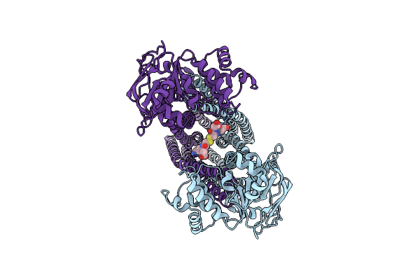
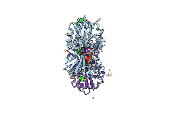
Organism: Alkaliphilus oremlandii
Method: X-RAY DIFFRACTION Resolution:1.95 Å Release Date: 2014-10-01 Classification: OXIDOREDUCTASE Ligands: ACT, GDS |
 |
Organism: Novosphingobium aromaticivorans
Method: X-RAY DIFFRACTION Resolution:2.35 Å Release Date: 2014-03-19 Classification: TRANSPORT PROTEIN Ligands: LDA, GDS, PO4 |
 |
Crystal Structure Of Glutathione Transferase Pput_1760 From Pseudomonas Putida, Target Efi-507288, With One Glutathione Disulfide Bound Per One Protein Subunit
Organism: Pseudomonas putida
Method: X-RAY DIFFRACTION Resolution:1.30 Å Release Date: 2013-11-06 Classification: TRANSFERASE Ligands: GDS, GOL, FMT |
 |
Crystal Structure Of Nu-Class Glutathione Transferase Yghu From Streptococcus Sanguinis Sk36, Complex With Glutathione Disulfide, Target Efi-507286
Organism: Streptococcus sanguinis
Method: X-RAY DIFFRACTION Resolution:1.95 Å Release Date: 2013-10-16 Classification: TRANSFERASE Ligands: GDS, ACT |
 |
Molecular And Structural Basis Of Glutathione Import In Gram-Positive Bacteria Via Gsht And The Cystine Abc Importer Tcybc Of Streptococcus Mutans.
Organism: Streptococcus mutans
Method: X-RAY DIFFRACTION Resolution:1.55 Å Release Date: 2013-08-21 Classification: TRANSPORT PROTEIN Ligands: GDS, CD, CL |
 |
Crystal Structure Of The Glutathione Transferase Ure2P1 From Phanerochaete Chrysosporium.
Organism: Phanerochaete chrysosporium
Method: X-RAY DIFFRACTION Resolution:1.45 Å Release Date: 2013-06-12 Classification: TRANSFERASE Ligands: GDS, GOL |
 |
Crystal Structure Of The Glutathione Transferase Ure2P5 From Phanerochaete Chrysosporium
Organism: Phanerochaete chrysosporium
Method: X-RAY DIFFRACTION Resolution:1.90 Å Release Date: 2013-06-12 Classification: TRANSFERASE Ligands: GDS, SO4, GOL |