Planned Maintenance: Some services may turn out to be unavailable from 15th January, 2026 to 16th January, 2026. We apologize for the inconvenience!

Planned Maintenance: Some services may turn out to be unavailable from 15th January, 2026 to 16th January, 2026. We apologize for the inconvenience!
 |
Organism: Homo sapiens
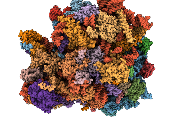
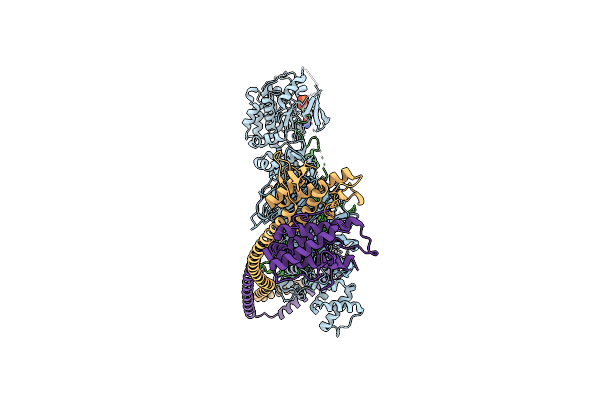
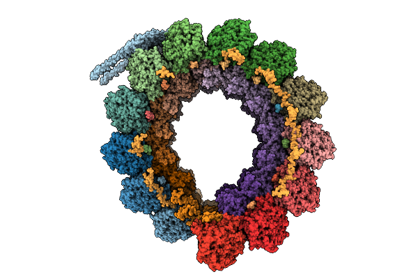
Method: ELECTRON MICROSCOPY Release Date: 2026-01-14 Classification: RIBOSOME Ligands: GDP, MG, K, ZN, PUT, HYG, SPD, 3HE, ANM, NA |
 |
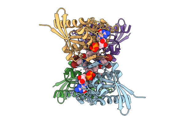
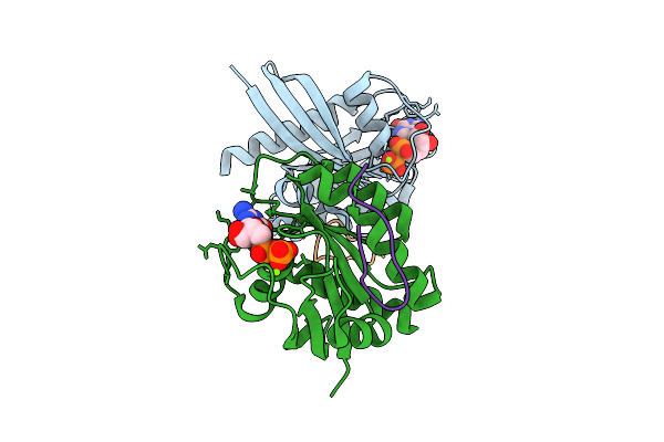
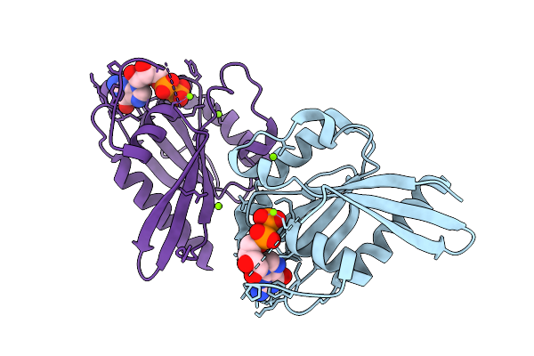
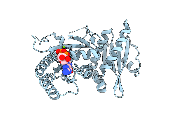
Crystal Structure Of Human Rab43 In Gdp-Alf3 Transition State Complex With Usp6Nl Tbc Domain
Organism: Homo sapiens
Method: X-RAY DIFFRACTION Release Date: 2026-01-14 Classification: HYDROLASE Ligands: MG, GDP, AF3, PEG |
 |
Organism: Mus musculus
Method: X-RAY DIFFRACTION Release Date: 2026-01-14 Classification: LIPID BINDING PROTEIN Ligands: GDP, GOL |
 |
Organism: Mus musculus
Method: X-RAY DIFFRACTION Release Date: 2026-01-14 Classification: LIPID BINDING PROTEIN Ligands: GDP |
 |
Organism: Mus musculus
Method: X-RAY DIFFRACTION Release Date: 2026-01-14 Classification: LIPID BINDING PROTEIN Ligands: GDP |
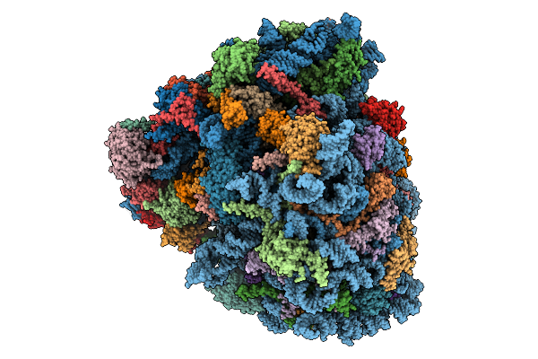 |
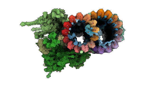
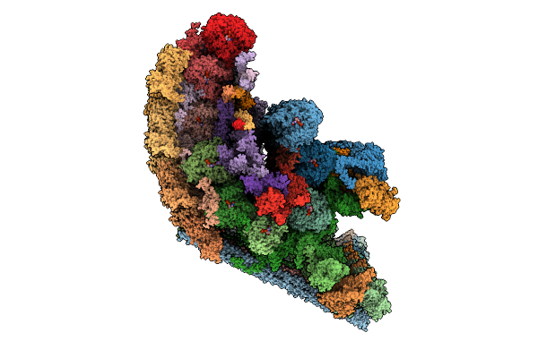
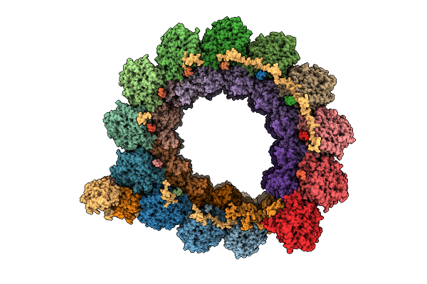
Gtpbp1*Gdp*Phe-Trna*Ribosome In The Post-Gtp Hydrolysis State, Structure Iv
Organism: Homo sapiens, Oryctolagus cuniculus, Saccharomyces cerevisiae
Method: ELECTRON MICROSCOPY Release Date: 2025-12-31 Classification: RIBOSOME Ligands: SPD, ZN, GTP, PHE, ATP, MET, K, GDP, MG |
 |
Organism: Homo sapiens
Method: X-RAY DIFFRACTION Release Date: 2025-12-24 Classification: HYDROLASE Ligands: GDP, EDO, A1IWE |
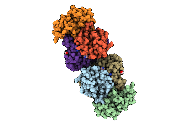 |
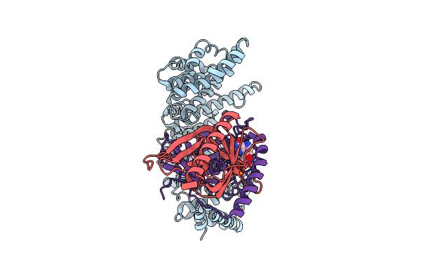
Rhs2-Ct Endonuclease Toxin In Complex With Cognate Immunity Protein Rhsi2 And Ef-Tu
Organism: Escherichia coli, Serratia marcescens
Method: X-RAY DIFFRACTION Release Date: 2025-12-24 Classification: ANTIMICROBIAL PROTEIN Ligands: GDP, EDO, MG, IMD |
 |
Organism: Homo sapiens
Method: ELECTRON MICROSCOPY Release Date: 2025-12-24 Classification: SIGNALING PROTEIN Ligands: GDP, MG, ZN |
 |
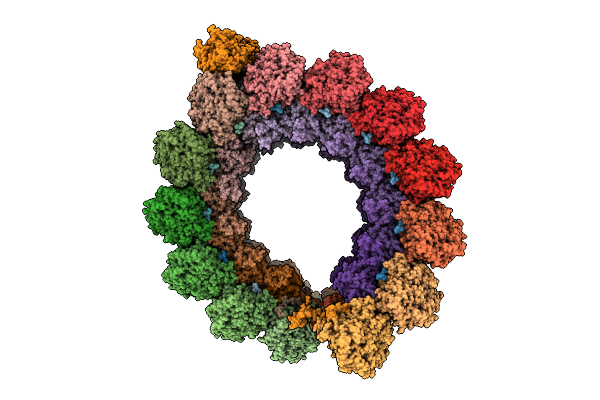
Organism: Leishmania tarentolae, Leishmania mexicana
Method: ELECTRON MICROSCOPY Release Date: 2025-12-17 Classification: STRUCTURAL PROTEIN Ligands: GDP, MG, GTP, ZN, CA, ATP |
 |
Organism: Homo sapiens, Synthetic construct
Method: X-RAY DIFFRACTION Release Date: 2025-12-10 Classification: HYDROLASE Ligands: MG, GDP |
 |
Organism: Homo sapiens
Method: X-RAY DIFFRACTION Release Date: 2025-12-10 Classification: ONCOPROTEIN Ligands: GDP, MG, EDO, A1BV7 |
 |
Organism: Bombyx mori
Method: X-RAY DIFFRACTION Release Date: 2025-12-10 Classification: PROTEIN BINDING Ligands: GDP, MG |
 |
Organism: Homo sapiens
Method: ELECTRON MICROSCOPY Release Date: 2025-12-10 Classification: TRANSPORT PROTEIN Ligands: MG, GDP |
 |
Organism: Homo sapiens
Method: X-RAY DIFFRACTION Release Date: 2025-12-03 Classification: ONCOPROTEIN/INHIBITOR Ligands: GDP, MG, A1CG4 |
 |
Organism: Toxoplasma gondii
Method: ELECTRON MICROSCOPY Release Date: 2025-12-03 Classification: STRUCTURAL PROTEIN Ligands: GTP, MG, GDP |
 |
Cryo-Em Structure Of Intraconoidal Microtubule 2 (Icmt2) From Toxoplasma Gondii (8-Nm Repeat)
Organism: Toxoplasma gondii
Method: ELECTRON MICROSCOPY Release Date: 2025-12-03 Classification: STRUCTURAL PROTEIN Ligands: GTP, MG, GDP |
 |
Cryo-Em Structure Of Intraconoidal Microtubule 1 (Icmt1) From Toxoplasma Gondii (8-Nm Repeat)
Organism: Toxoplasma gondii
Method: ELECTRON MICROSCOPY Release Date: 2025-12-03 Classification: STRUCTURAL PROTEIN Ligands: GTP, MG, GDP |
 |
Cryo-Em Structure Of The Apical Region Of Subpellicular Microtubule (Spmt) From Toxoplasma Gondii (8-Nm Repeat)
Organism: Toxoplasma gondii
Method: ELECTRON MICROSCOPY Release Date: 2025-12-03 Classification: STRUCTURAL PROTEIN Ligands: GDP, GTP, MG |
 |
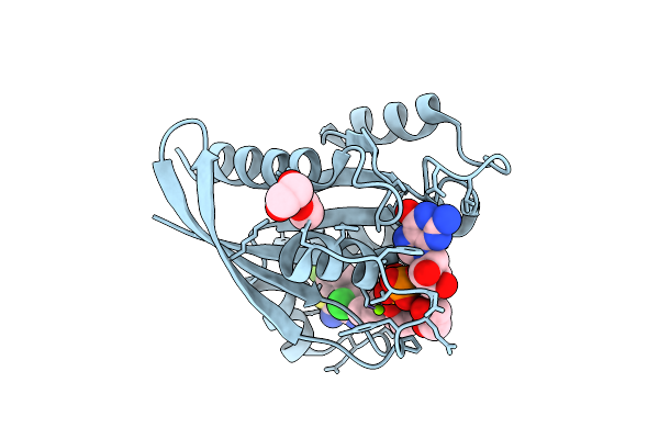
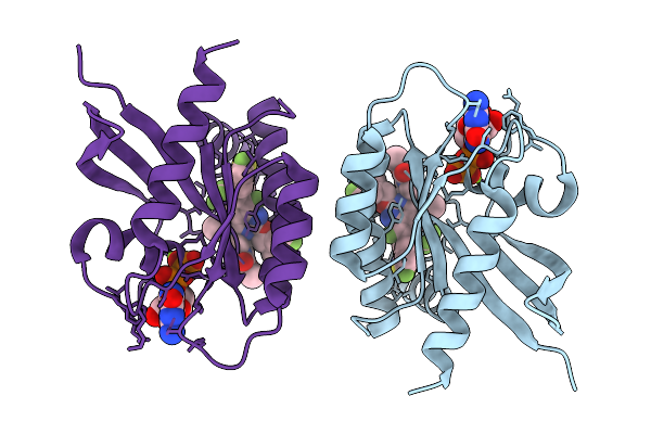
Crystal Structure Of Monomeric Rag-Like Small Gtpase From Asgard Lokiarchaeota (Lokiragm) In Complex With Gdp
Organism: Candidatus prometheoarchaeum syntrophicum
Method: X-RAY DIFFRACTION Release Date: 2025-11-26 Classification: HYDROLASE Ligands: GDP, MG |