
Search Count: 93
 |
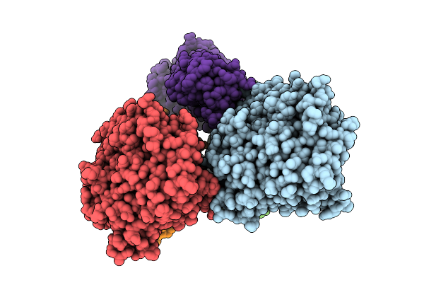
Organism: Goslarvirus
Method: ELECTRON MICROSCOPY Release Date: 2025-12-24 Classification: VIRAL PROTEIN Ligands: G2P |
 |
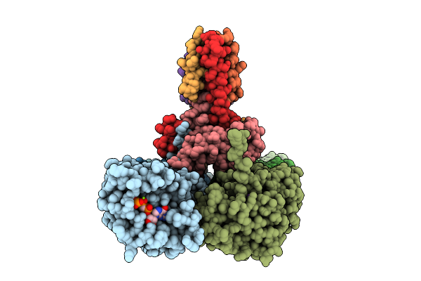
Organism: Goslarvirus
Method: ELECTRON MICROSCOPY Release Date: 2025-12-24 Classification: VIRAL PROTEIN Ligands: G2P |
 |
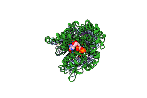
Organism: Thermus thermophilus hb8, Synthetic construct
Method: ELECTRON MICROSCOPY Release Date: 2025-12-17 Classification: TRANSCRIPTION Ligands: MG, B4P, ZN, G2P |
 |
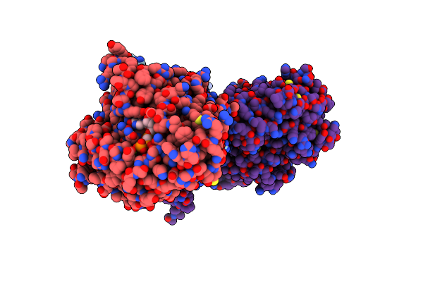
Extended, Cyr715-Bound State Of Manduca Sexta Soluble Guanylate Cyclase Mutant Beta C122S
Organism: Manduca sexta
Method: ELECTRON MICROSCOPY Release Date: 2025-11-12 Classification: SIGNALING PROTEIN Ligands: G2P, HEM, A1CGK |
 |
Organism: Xenopus laevis, Bos taurus
Method: ELECTRON MICROSCOPY Release Date: 2025-11-05 Classification: CELL CYCLE Ligands: G2P, MG, GTP |
 |
Organism: Pseudomonas pavonaceae, Homo sapiens
Method: ELECTRON MICROSCOPY Release Date: 2025-09-17 Classification: LIGASE Ligands: G2P, MG, TA1 |
 |
Organism: Xenopus laevis, Bos taurus
Method: ELECTRON MICROSCOPY Release Date: 2025-08-13 Classification: CELL CYCLE Ligands: GTP, MG, G2P |
 |
Organism: Candidatus odinarchaeota
Method: ELECTRON MICROSCOPY Release Date: 2025-07-30 Classification: CELL CYCLE Ligands: G2P |
 |
Organism: Klebsiella pneumoniae subsp. pneumoniae mgh 78578, Klebsiella pneumoniae 342
Method: ELECTRON MICROSCOPY Release Date: 2025-07-23 Classification: CELL CYCLE Ligands: G2P, MG, K |
 |
Organism: Homo sapiens, Mus musculus
Method: ELECTRON MICROSCOPY Release Date: 2024-11-27 Classification: LYASE Ligands: G2P, MG |
 |
Organism: Homo sapiens, Sus scrofa
Method: ELECTRON MICROSCOPY Release Date: 2024-11-27 Classification: CELL CYCLE Ligands: G2P, MG, GTP |
 |
Organism: Caenorhabditis elegans
Method: ELECTRON MICROSCOPY Release Date: 2024-11-06 Classification: STRUCTURAL PROTEIN Ligands: GTP, G2P |
 |
Organism: Caenorhabditis elegans
Method: ELECTRON MICROSCOPY Release Date: 2024-11-06 Classification: STRUCTURAL PROTEIN Ligands: GTP, PIN, G2P |
 |
Organism: Caenorhabditis elegans
Method: ELECTRON MICROSCOPY Release Date: 2024-11-06 Classification: STRUCTURAL PROTEIN Ligands: GTP, ACO, G2P |
 |
Organism: Caenorhabditis elegans
Method: ELECTRON MICROSCOPY Release Date: 2024-11-06 Classification: STRUCTURAL PROTEIN Ligands: GTP, ACO, G2P |
 |
Organism: Homo sapiens, Sus scrofa
Method: ELECTRON MICROSCOPY Release Date: 2024-07-17 Classification: HYDROLASE/SUBSTRATE,STRUCTURAL PROTEIN Ligands: MG, GTP, G2P, ZN, GLU |
 |
Organism: Homo sapiens, Sus scrofa
Method: ELECTRON MICROSCOPY Release Date: 2024-07-17 Classification: HYDROLASE/SUBSTRATE,STRUCTURAL PROTEIN Ligands: MG, GTP, G2P, GLU, ZN |
 |
Organism: Homo sapiens, Sus scrofa
Method: ELECTRON MICROSCOPY Release Date: 2024-07-17 Classification: HYDROLASE/SUBSTRATE,STRUCTURAL PROTEIN Ligands: MG, GTP, G2P, GLU, ZN |
 |
Organism: Homo sapiens
Method: ELECTRON MICROSCOPY Release Date: 2024-06-12 Classification: STRUCTURAL PROTEIN Ligands: GTP, MG, G2P |
 |
Organism: Mus musculus, Homo sapiens
Method: ELECTRON MICROSCOPY Release Date: 2024-05-29 Classification: LIGASE Ligands: GTP, MG, G2P, ATP |