
Search Count: 11
 |
Organism: Hepatitis b virus subtype adyw
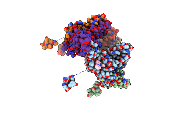
Method: X-RAY DIFFRACTION Resolution:2.60 Å Release Date: 2024-09-11 Classification: VIRUS LIKE PARTICLE Ligands: 5PT |
 |
Organism: Guillardia theta ccmp2712
Method: ELECTRON MICROSCOPY Resolution:2.86 Å Release Date: 2024-09-04 Classification: MEMBRANE PROTEIN Ligands: RET |
 |
Organism: Guillardia theta ccmp2712
Method: ELECTRON MICROSCOPY Resolution:2.73 Å Release Date: 2024-09-04 Classification: MEMBRANE PROTEIN Ligands: RET, PC1 |
 |
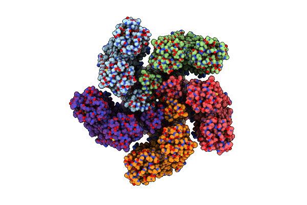
Organism: Guillardia theta
Method: ELECTRON MICROSCOPY Resolution:2.71 Å Release Date: 2024-09-04 Classification: MEMBRANE PROTEIN Ligands: RET, PC1 |
 |
Cryo-Em Structure Of Bestrhodopsin (Rhodopsin-Rhodopsin-Bestrophin) Complex
Organism: Phaeocystis
Method: ELECTRON MICROSCOPY Release Date: 2022-07-06 Classification: MEMBRANE PROTEIN Ligands: RET |
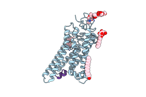 |
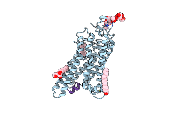
Organism: Bos taurus
Method: X-RAY DIFFRACTION Resolution:2.91 Å Release Date: 2020-07-01 Classification: SIGNALING PROTEIN Ligands: BOG, PLM, 64Z |
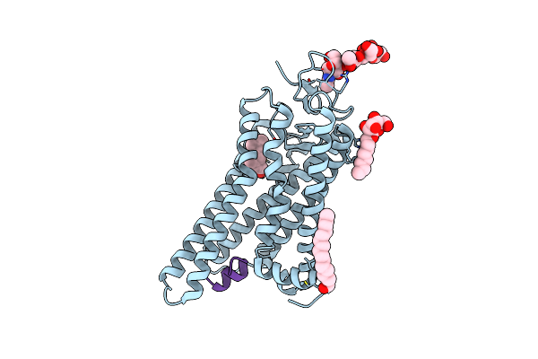 |
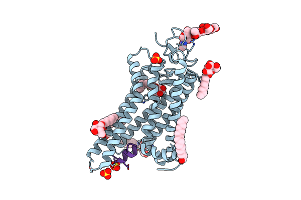
Organism: Bos taurus
Method: X-RAY DIFFRACTION Resolution:2.90 Å Release Date: 2020-07-01 Classification: SIGNALING PROTEIN Ligands: BOG, PLM, NZZ |
 |
Organism: Bos taurus
Method: X-RAY DIFFRACTION Resolution:3.19 Å Release Date: 2020-06-24 Classification: SIGNALING PROTEIN Ligands: ODM, BOG, PLM |
 |
Organism: Bos taurus
Method: X-RAY DIFFRACTION Resolution:2.71 Å Release Date: 2020-02-12 Classification: SIGNALING PROTEIN Ligands: BOG, PLM, SO4 |
 |
Organism: Thermoplasmatales archaeon sg8-52-1
Method: X-RAY DIFFRACTION Resolution:2.40 Å Release Date: 2019-09-25 Classification: MEMBRANE PROTEIN Ligands: OLC, RET |
 |
Crystal Structure Of The P2X4 Receptor From Zebrafish In The Presence Of Ctp At 2.8 Angstroms
Organism: Danio rerio
Method: X-RAY DIFFRACTION Resolution:2.80 Å Release Date: 2017-04-05 Classification: MEMBRANE PROTEIN Ligands: NAG, GOL, CTP |