
Search Count: 6
 |
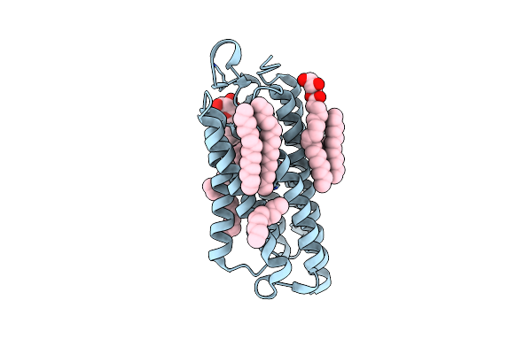
Crystal Structure Of The Cell-Free Synthesized Membrane Protein, Acetabularia Rhodopsin I, At 1.57 Angstrom
Organism: Acetabularia acetabulum
Method: X-RAY DIFFRACTION Resolution:1.57 Å Release Date: 2015-08-26 Classification: PROTON TRANSPORT Ligands: RET, OLB, D12, D10, OCT, C14 |
 |
Crystal Structure Of The Cell-Free Synthesized Membrane Protein, Acetabularia Rhodopsin I, At 1.52 Angstrom
Organism: Acetabularia acetabulum
Method: X-RAY DIFFRACTION Resolution:1.52 Å Release Date: 2015-08-26 Classification: PROTON TRANSPORT Ligands: RET, OLB, D12, C14, R16, D10 |
 |
Crystal Structure Of The Cell-Free Synthesized Membrane Protein, Acetabularia Rhodopsin I, At 1.80 Angstrom
Organism: Acetabularia acetabulum
Method: X-RAY DIFFRACTION Resolution:1.80 Å Release Date: 2015-08-26 Classification: PROTON TRANSPORT Ligands: RET, OLB, C14, D10, R16, D12 |
 |
Crystal Structure Analysis Of The Pdz Domain Derived From The Tight Junction Regulating Protein
Organism: Mus musculus
Method: X-RAY DIFFRACTION Resolution:1.20 Å Release Date: 2013-03-27 Classification: PEPTIDE BINDING PROTEIN |
 |
Crystal Structure Analysis Of The Pdz Domain Derived From The Tight Junction Regulating Protein
Organism: Mus musculus
Method: X-RAY DIFFRACTION Resolution:1.35 Å Release Date: 2013-03-27 Classification: PEPTIDE BINDING PROTEIN Ligands: SO4 |
 |
Interplay Between Phosphatidyl-Inositol-Phosphates And Claudins Upon Binding To The 1St Pdz Domain Of Zonula Occludens 1
|