
Search Count: 16
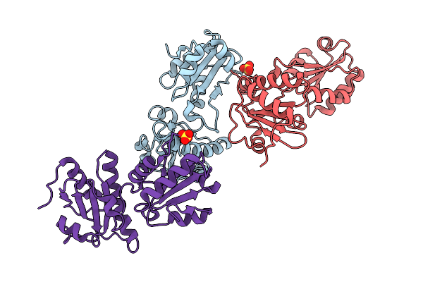 |
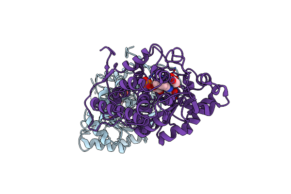
Organism: Homo sapiens
Method: X-RAY DIFFRACTION Release Date: 2025-10-01 Classification: ISOMERASE Ligands: SO4 |
 |
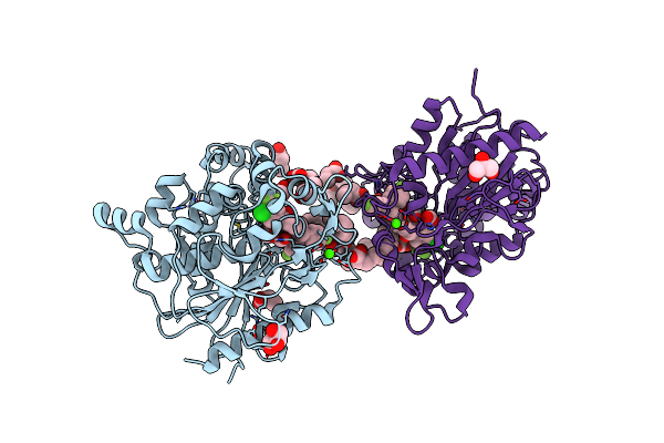 |
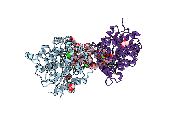
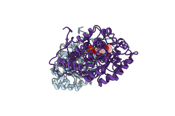
Organism: Staphylococcus aureus
Method: X-RAY DIFFRACTION Resolution:2.57 Å Release Date: 2025-04-30 Classification: HYDROLASE Ligands: OCA, PPI, GOL, A1L60, FMT, ZN, CA, 6NA, CL, BUA, 11A |
 |
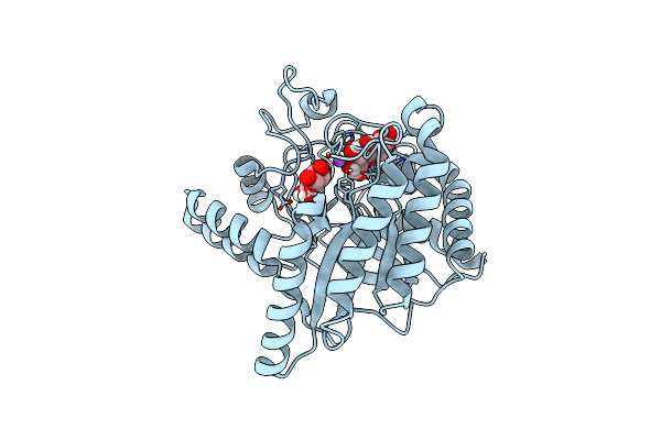
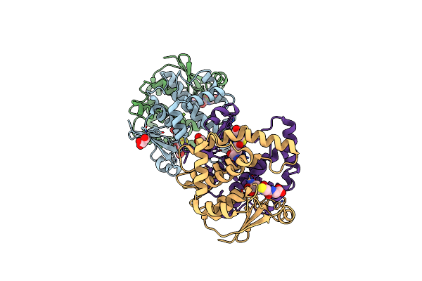
Neutron Structure Of Cellulase Cel6A From Phanerochaete Chrysosporium At Room Temperature
Organism: Phanerodontia chrysosporium
Method: X-RAY DIFFRACTION, NEUTRON DIFFRACTION Resolution:1.36 Å, 1.86 Å Release Date: 2025-03-12 Classification: HYDROLASE |
 |
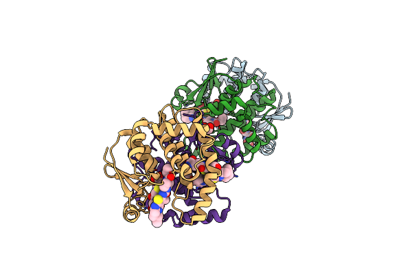
Neutron Structure Of Cellulase Cel6A From Phanerochaete Chrysosporium At Room Temperature, Enzyme-Product Complex
Organism: Phanerodontia chrysosporium
Method: X-RAY DIFFRACTION, NEUTRON DIFFRACTION Resolution:1.40 Å, 1.8 Å Release Date: 2025-03-12 Classification: HYDROLASE Ligands: BGC, NA |
 |
Neutron Structure Of Cellulase Cel6A From Phanerochaete Chrysosporium At Room Temperature, Low-D2O-Solvent
Organism: Phanerodontia chrysosporium
Method: X-RAY DIFFRACTION, NEUTRON DIFFRACTION Resolution:1.40 Å, 2.15 Å Release Date: 2025-03-12 Classification: HYDROLASE |
 |
Neutron Structure Of Cellulase Cel6A From Phanerochaete Chrysosporium At Room Temperature, Enzyme-Product Complex, H2O Solvent
Organism: Phanerodontia chrysosporium
Method: X-RAY DIFFRACTION, NEUTRON DIFFRACTION Resolution:1.80 Å, 2.59 Å Release Date: 2025-03-12 Classification: HYDROLASE Ligands: BGC, NA |
 |
High-Resolution X-Ray Structure Of Cellulase Cel6A From Phanerochaete Chrysosporium At Cryogenic Temperature
Organism: Phanerodontia chrysosporium
Method: X-RAY DIFFRACTION Resolution:0.80 Å Release Date: 2025-03-12 Classification: HYDROLASE Ligands: MPD, ACT |
 |
High-Resolution X-Ray Structure Of Cellulase Cel6A From Phanerochaete Chrysosporium At Cryogenic Temperature, Enzyme-Product Complex
Organism: Phanerodontia chrysosporium
Method: X-RAY DIFFRACTION Resolution:0.85 Å Release Date: 2025-03-12 Classification: HYDROLASE Ligands: BGC, NA, CL, PEG |
 |
Organism: Aerococcus viridans
Method: X-RAY DIFFRACTION Resolution:1.33 Å Release Date: 2022-06-15 Classification: OXIDOREDUCTASE Ligands: ACT, FMN, EDO |
 |
Organism: Aerococcus viridans
Method: X-RAY DIFFRACTION Resolution:1.30 Å Release Date: 2022-03-23 Classification: OXIDOREDUCTASE Ligands: FMN, 2OP |
 |
Organism: Aerococcus viridans
Method: X-RAY DIFFRACTION Resolution:1.38 Å Release Date: 2022-03-23 Classification: OXIDOREDUCTASE Ligands: FMN, LAC |
 |
Organism: Aerococcus viridans
Method: X-RAY DIFFRACTION Resolution:1.41 Å Release Date: 2022-03-23 Classification: OXIDOREDUCTASE Ligands: FMN, PYR |
 |
Organism: Homo sapiens
Method: X-RAY DIFFRACTION Resolution:1.68 Å Release Date: 2018-12-05 Classification: TRANSFERASE Ligands: GSH, GOL, MG |
 |
Crystal Structure Of Hematopoietic Prostaglandin D Synthase In Complex With U004
Organism: Homo sapiens
Method: X-RAY DIFFRACTION Resolution:1.39 Å Release Date: 2018-12-05 Classification: TRANSFERASE/TRANSFERASE INHIBITOR Ligands: MG, GSH, UX4, GOL |
 |
Organism: Rattus norvegicus
Method: X-RAY DIFFRACTION Resolution:1.09 Å Release Date: 2018-09-19 Classification: TRANSFERASE Ligands: GSH, MG, DIO, EDO, GOL, PGE |