
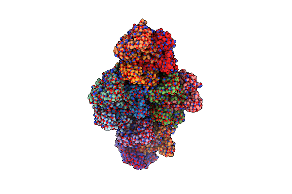
Search Count: 14
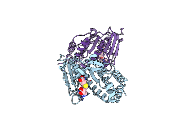 |
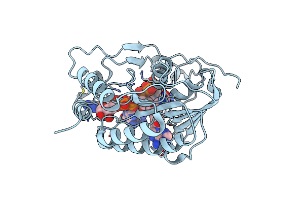
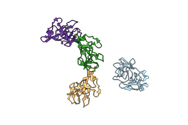
Crystal Structure Of The Murray Valley Encephalitis Virus Ns5 2'-O Methyltransferase Domain In Complex With Sah (Monoclinic Form 1)
Organism: Murray valley encephalitis virus (strain mve-1-51)
Method: X-RAY DIFFRACTION Resolution:2.00 Å Release Date: 2007-05-29 Classification: TRANSFERASE Ligands: CL, SAH |
 |
Crystal Structure Of The Murray Valley Encephalitis Virus Ns5 2'-O Methyltransferase Domain In Complex With Sah (Monoclinic Form 2)
Organism: Murray valley encephalitis virus (strain mve-1-51)
Method: X-RAY DIFFRACTION Resolution:2.20 Å Release Date: 2007-05-29 Classification: TRANSFERASE Ligands: CL, SO4, UNX, SAH, GOL |
 |
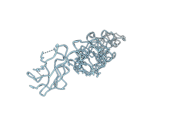
Crystal Structure Of The Murray Valley Encephalitis Virus Ns5 2'-O Methyltransferase Domain In Complex With Sah (Orthorhombic Crystal Form)
Organism: Murray valley encephalitis virus (strain mve-1-51)
Method: X-RAY DIFFRACTION Resolution:2.30 Å Release Date: 2007-05-29 Classification: TRANSFERASE Ligands: SO4, SAH |
 |
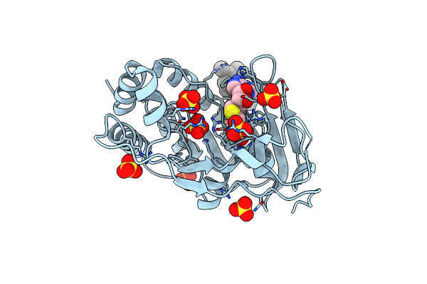
Crystal Structure Of The Murray Valley Encephalitis Virus Ns5 2'-O Methyltransferase Domain In Complex With Sah And 7M-Gtp
Organism: Murray valley encephalitis virus (strain mve-1-51)
Method: X-RAY DIFFRACTION Resolution:2.20 Å Release Date: 2007-05-29 Classification: TRANSFERASE Ligands: MGT, 3PO, SAH, GOL, CL |
 |
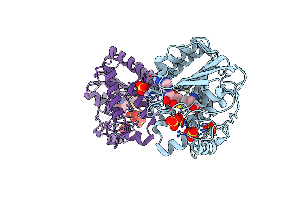
Crystal Structure Of The Murray Valley Encephalitis Virus Ns5 2'-O Methyltransferase Domain In Complex With Sah And Gtpg
Organism: Murray valley encephalitis virus (strain mve-1-51)
Method: X-RAY DIFFRACTION Resolution:2.30 Å Release Date: 2007-05-29 Classification: TRANSFERASE Ligands: GTP, SAH |
 |
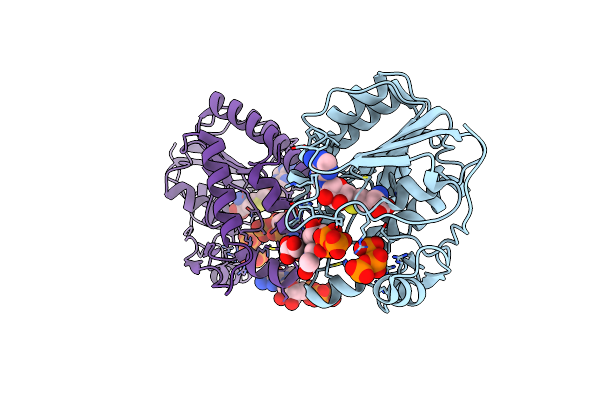
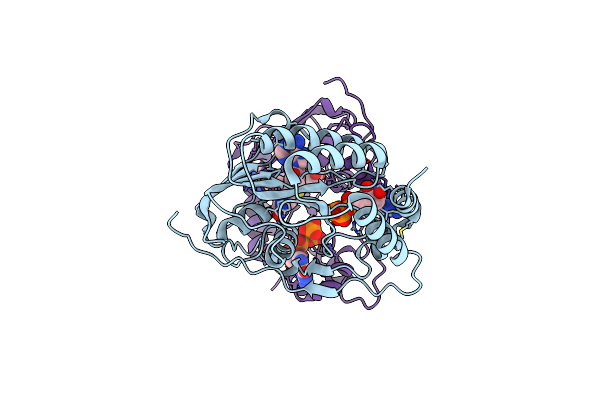
Crystal Structure Of The Murray Valley Encephalitis Virus Ns5 2'-O Methyltransferase Domain In Complex With Sam And Gtpa
Organism: Murray valley encephalitis virus (strain mve-1-51)
Method: X-RAY DIFFRACTION Resolution:2.80 Å Release Date: 2007-05-29 Classification: TRANSFERASE Ligands: SAM, G3A |
 |
Organism: Enterobacteria phage prd1
Method: X-RAY DIFFRACTION Resolution:4.20 Å Release Date: 2004-11-11 Classification: VIRUS |
 |
Organism: Semliki forest virus
Method: X-RAY DIFFRACTION Resolution:3.00 Å Release Date: 2002-04-06 Classification: VIRAL PROTEIN |
 |
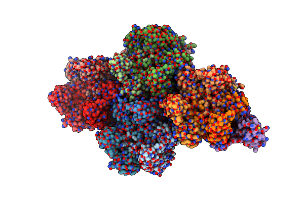
Quasi-Atomic Resolution Model Of Bacteriophage Prd1 Sus607 Mutant, Obtained By Combined Cryo-Em And X-Ray Crystallography.
Organism: Bacteriophage prd1
Method: ELECTRON MICROSCOPY Resolution:13.30 Å Release Date: 2002-03-15 Classification: VIRUS/VIRAL PROTEIN |
 |
Quasi-Atomic Resolution Model Of Bacteriophage Prd1 Capsid, Obtained By Combined Cryo-Em And X-Ray Crystallography.
Organism: Bacteriophage prd1
Method: ELECTRON MICROSCOPY Resolution:13.50 Å Release Date: 2002-03-13 Classification: VIRUS/VIRAL PROTEIN |
 |
Quasi-Atomic Resolution Model Of Bacteriophage Prd1 P3-Shell, Obtained By Combined Cryo-Em And X-Ray Crystallography.
Organism: Bacteriophage prd1
Method: ELECTRON MICROSCOPY Resolution:12.00 Å Release Date: 2001-12-05 Classification: VIRUS |
 |
Quasi-Atomic Resolution Model Of Bacteriophage Prd1 Sus1 Mutant, Obtained By Combined Cryo-Em And X-Ray Crystallography.
Organism: Bacteriophage prd1
Method: ELECTRON MICROSCOPY Resolution:14.00 Å Release Date: 2001-12-05 Classification: VIRUS |
 |
Quasi-Atomic Resolution Model Of Bacteriophage Prd1 Wild Type Virion, Obtained By Combined Cryo-Em And X-Ray Crystallography.
Organism: Bacteriophage prd1
Method: ELECTRON MICROSCOPY Resolution:25.00 Å Release Date: 2001-12-05 Classification: VIRUS |
 |
9 Angstrom Resolution Cryo-Em Reconstruction Structure Of Semliki Forest Virus (Sfv) And Fitting Of The Capsid Protein Structure In The Em Density
Organism: Semliki forest virus
Method: ELECTRON MICROSCOPY Resolution:9.00 Å Release Date: 2000-08-18 Classification: VIRUS/VIRAL PROTEIN |