Search Count: 52
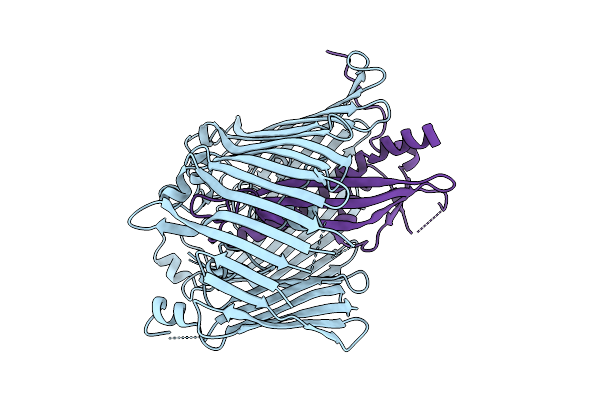 |
Organism: Escherichia coli
Method: ELECTRON MICROSCOPY Release Date: 2025-12-10 Classification: TRANSPORT PROTEIN |
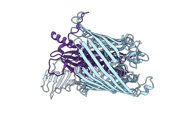 |
Organism: Escherichia coli
Method: ELECTRON MICROSCOPY Release Date: 2025-12-10 Classification: TRANSPORT PROTEIN |
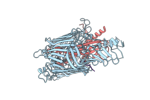 |
Organism: Escherichia coli
Method: ELECTRON MICROSCOPY Release Date: 2025-12-10 Classification: TRANSPORT PROTEIN |
 |
Organism: Escherichia coli
Method: ELECTRON MICROSCOPY Release Date: 2025-12-10 Classification: TRANSPORT PROTEIN |
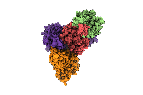 |
Organism: Bacillus subtilis subsp. subtilis str. 168
Method: ELECTRON MICROSCOPY Release Date: 2025-11-26 Classification: PROTEIN TRANSPORT |
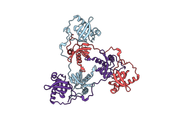 |
Crystal Structure Of A Double Mutant Of Virb8-Like Orfg Central And C-Terminal Domains Of Streptococcus Thermophilus Icest3 (Gram Positive Conjugative Type Iv Secretion System).
Organism: Streptococcus thermophilus
Method: X-RAY DIFFRACTION Resolution:2.60 Å Release Date: 2025-03-12 Classification: TRANSPORT PROTEIN |
 |
Organism: Streptococcus pneumoniae, Synthetic construct
Method: ELECTRON MICROSCOPY Release Date: 2024-10-30 Classification: DNA BINDING PROTEIN Ligands: AGS, MG |
 |
Organism: Legionella pneumophila, Synthetic construct
Method: ELECTRON MICROSCOPY Release Date: 2024-10-30 Classification: DNA BINDING PROTEIN Ligands: ANP |
 |
Organism: Legionella pneumophila, Synthetic construct
Method: ELECTRON MICROSCOPY Release Date: 2024-10-30 Classification: DNA BINDING PROTEIN Ligands: ANP |
 |
Organism: Legionella pneumophila
Method: ELECTRON MICROSCOPY Release Date: 2024-10-30 Classification: DNA BINDING PROTEIN |
 |
Organism: Legionella pneumophila
Method: ELECTRON MICROSCOPY Release Date: 2024-10-30 Classification: DNA BINDING PROTEIN Ligands: ANP |
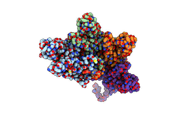 |
Organism: Streptococcus pneumoniae, Lambdavirus lambda
Method: ELECTRON MICROSCOPY Release Date: 2024-02-21 Classification: RECOMBINATION Ligands: AGS |
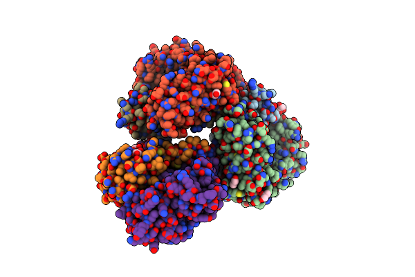 |
Organism: Drosophila melanogaster
Method: X-RAY DIFFRACTION Resolution:2.10 Å Release Date: 2024-02-21 Classification: SIGNALING PROTEIN Ligands: GOL |
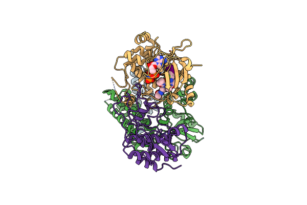 |
Organism: Drosophila melanogaster
Method: ELECTRON MICROSCOPY Release Date: 2024-02-21 Classification: SIGNALING PROTEIN Ligands: ANP, QOM, MG |
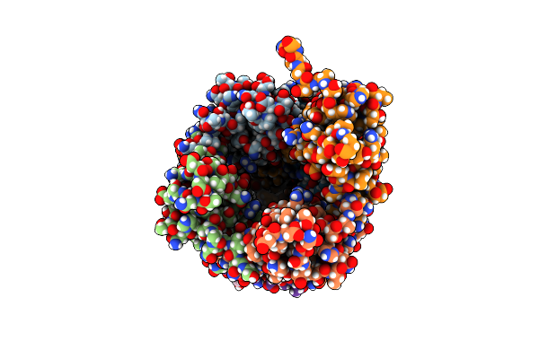 |
Organism: Streptococcus sanguinis
Method: ELECTRON MICROSCOPY Release Date: 2023-11-22 Classification: PROTEIN FIBRIL |
 |
Organism: Synthetic construct, Helicobacter pylori 26695
Method: X-RAY DIFFRACTION Resolution:2.00 Å Release Date: 2023-05-17 Classification: TOXIN |
 |
Organism: Synthetic construct, Helicobacter pylori 26695
Method: X-RAY DIFFRACTION Resolution:1.84 Å Release Date: 2023-05-17 Classification: TOXIN |
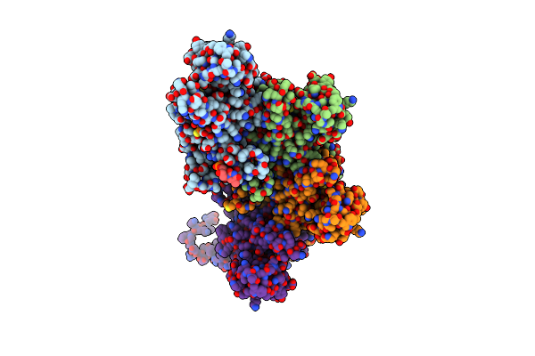 |
Organism: Streptococcus pneumoniae, Bacteriophage sp.
Method: ELECTRON MICROSCOPY Release Date: 2023-03-22 Classification: RECOMBINATION Ligands: AGS |
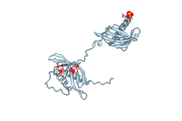 |
Crystal Structure Of Virb8-Like Orfg Central And C-Terminal Domains Of Streptococcus Thermophilus Icest3 (Gram Positive Conjugative Type Iv Secretion System).
Organism: Streptococcus thermophilus
Method: X-RAY DIFFRACTION Resolution:1.84 Å Release Date: 2022-09-07 Classification: TRANSPORT PROTEIN Ligands: SO4, GOL |
 |
Photorhabdus Laumondii T6Ss-Associated Rhs Protein Carrying The Tre23 Toxin Domain
Organism: Photorhabdus laumondii subsp. laumondii tto1
Method: ELECTRON MICROSCOPY Resolution:3.17 Å Release Date: 2021-12-01 Classification: TOXIN |

