
Search Count: 91
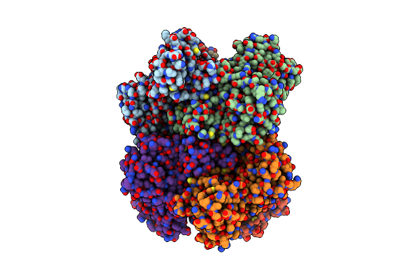 |
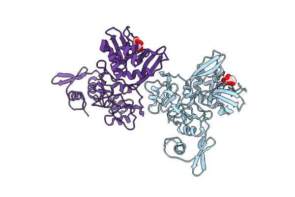
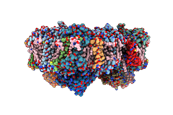
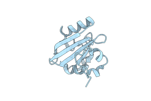
Structural Asymmetry In Sars-Cov-2 Nsp15 Hexamer Important For Catalytic Activity
Organism: Severe acute respiratory syndrome coronavirus 2
Method: X-RAY DIFFRACTION Release Date: 2025-06-04 Classification: VIRAL PROTEIN |
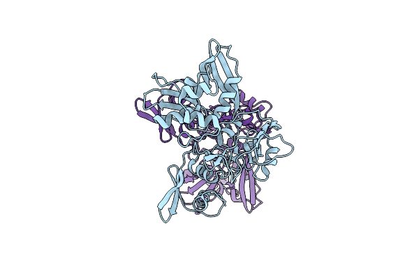 |
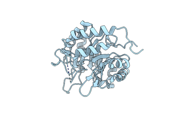
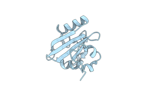
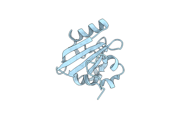
Organism: Severe acute respiratory syndrome coronavirus 2
Method: X-RAY DIFFRACTION Resolution:3.00 Å Release Date: 2025-06-04 Classification: VIRAL PROTEIN |
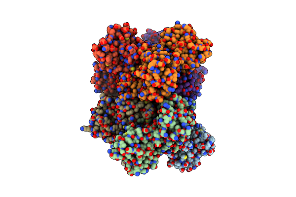 |
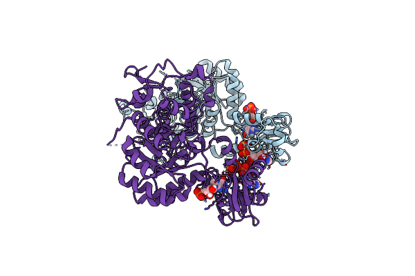
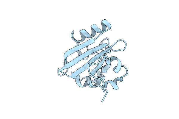
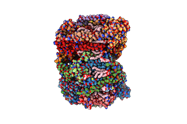
Organism: Severe acute respiratory syndrome coronavirus 2
Method: X-RAY DIFFRACTION Resolution:3.00 Å Release Date: 2025-06-04 Classification: VIRAL PROTEIN |
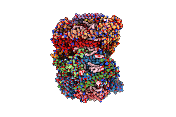 |
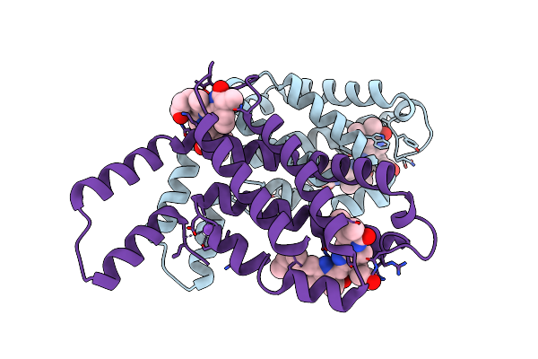
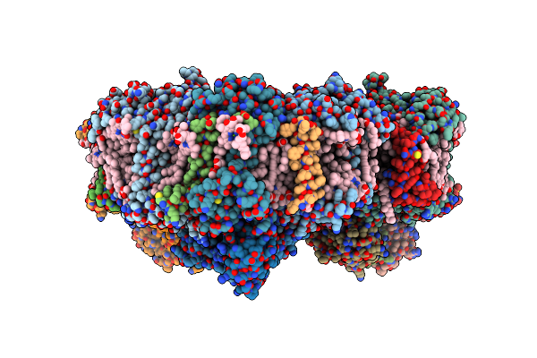
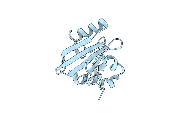
Time-Resolve Sfx Structure Of A Photoproduct Of Carbon Monoxide Complex Of Bovine Cytochrome C Oxidase
Organism: Bos taurus
Method: X-RAY DIFFRACTION Resolution:2.80 Å Release Date: 2023-09-20 Classification: MEMBRANE PROTEIN Ligands: HEA, CU, MG, NA, TGL, PGV, CMO, CUA, CHD, OH, CDL, PSC, ZN, PEK, DMU, SAC |
 |
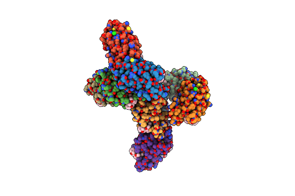
Organism: Homo sapiens
Method: X-RAY DIFFRACTION Resolution:2.70 Å Release Date: 2023-06-28 Classification: OXIDOREDUCTASE Ligands: FAD, ACT |
 |
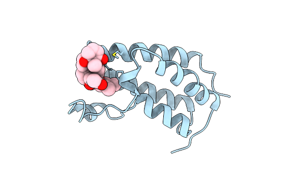
Phycocyanin Structure From A Modular Droplet Injector For Serial Femtosecond Crystallography
Organism: Thermosynechococcus vestitus bp-1
Method: X-RAY DIFFRACTION Resolution:2.00 Å Release Date: 2023-06-28 Classification: PLANT PROTEIN Ligands: CYC, NA |
 |
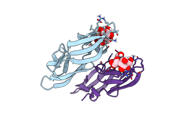
Organism: Homo sapiens
Method: X-RAY DIFFRACTION Resolution:2.89 Å Release Date: 2023-01-11 Classification: TRANSCRIPTION Ligands: 8L6, CL |
 |
Organism: Homo sapiens
Method: X-RAY DIFFRACTION Resolution:1.80 Å Release Date: 2022-09-21 Classification: SIGNALING PROTEIN Ligands: 8L6 |
 |
Organism: Nostoc ellipsosporum
Method: X-RAY DIFFRACTION Resolution:1.25 Å Release Date: 2021-06-02 Classification: ANTIVIRAL PROTEIN |
 |
Room Temperature Structure Of Nsp15 Endoribonuclease From Sars Cov-2 Solved Using Sfx.
Organism: Severe acute respiratory syndrome coronavirus 2
Method: X-RAY DIFFRACTION Resolution:2.60 Å Release Date: 2020-10-21 Classification: VIRAL PROTEIN Ligands: CIT |
 |
Organism: Escherichia coli (strain b / bl21-de3)
Method: X-RAY DIFFRACTION Resolution:2.80 Å Release Date: 2020-09-02 Classification: TRANSFERASE |
 |
In Cellulo Crystallization Of Trypanosoma Brucei Imp Dehydrogenase Enables The Identification Of Atp And Gmp As Genuine Co-Factors
Organism: Trypanosoma brucei brucei
Method: X-RAY DIFFRACTION Resolution:2.80 Å Release Date: 2020-02-19 Classification: IMMUNOSUPPRESSANT Ligands: ATP, 5GP |
 |
Membrane Protein Megahertz Crystallography At The European Xfel, Photosystem I Xfel At 2.9 A
Organism: Thermosynechococcus elongatus (strain bp-1)
Method: X-RAY DIFFRACTION Resolution:2.90 Å Release Date: 2019-11-27 Classification: PHOTOSYNTHESIS Ligands: CL0, CLA, PQN, SF4, BCR, LHG, LMG, CA |
 |
Membrane Protein Megahertz Crystallography At The European Xfel, Photosystem I At Synchrotron To 2.9 A
Organism: Thermosynechococcus elongatus (strain bp-1)
Method: X-RAY DIFFRACTION Resolution:2.90 Å Release Date: 2019-11-20 Classification: PHOTOSYNTHESIS Ligands: CL0, CLA, PQN, SF4, BCR, LHG, LMG, CA |
 |
Organism: Halorhodospira halophila
Method: X-RAY DIFFRACTION Resolution:1.60 Å Release Date: 2019-09-18 Classification: SIGNALING PROTEIN |
 |
Organism: Halorhodospira halophila
Method: X-RAY DIFFRACTION Resolution:1.60 Å Release Date: 2019-09-18 Classification: SIGNALING PROTEIN |
 |
Organism: Halorhodospira halophila
Method: X-RAY DIFFRACTION Resolution:1.60 Å Release Date: 2019-09-18 Classification: SIGNALING PROTEIN |
 |
Organism: Halorhodospira halophila
Method: X-RAY DIFFRACTION Resolution:1.70 Å Release Date: 2019-09-18 Classification: SIGNALING PROTEIN |
 |
Organism: Halorhodospira halophila
Method: X-RAY DIFFRACTION Resolution:1.60 Å Release Date: 2019-09-18 Classification: SIGNALING PROTEIN |
 |
Time-Resolved Sfx Structure Of The Pr Intermediate Of Cytochrome C Oxidase At Room Temperature
Organism: Bos taurus
Method: X-RAY DIFFRACTION Resolution:2.50 Å Release Date: 2019-03-20 Classification: OXIDOREDUCTASE Ligands: CU, MG, NA, HEA, PGV, TGL, O, OH, CUA, PSC, CHD, PEK, CDL, ZN, DMU |