
Search Count: 35
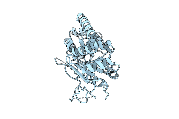 |
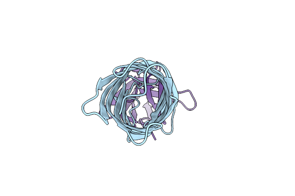
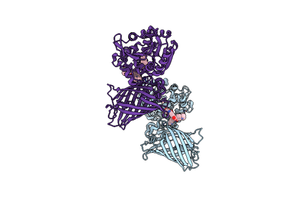
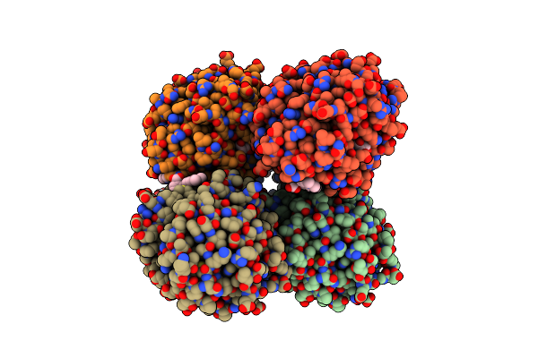
Organism: Homo sapiens
Method: X-RAY DIFFRACTION Resolution:2.15 Å Release Date: 2025-08-20 Classification: HYDROLASE Ligands: ZN |
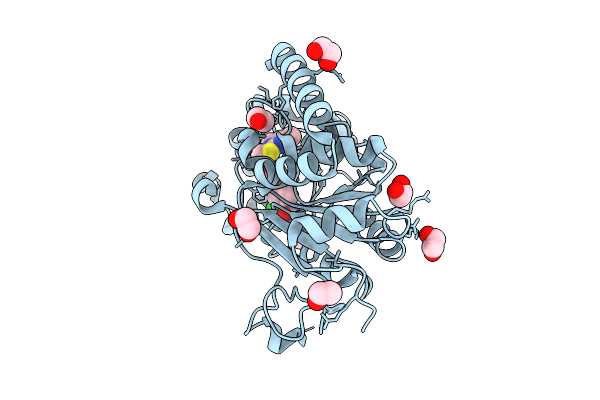 |
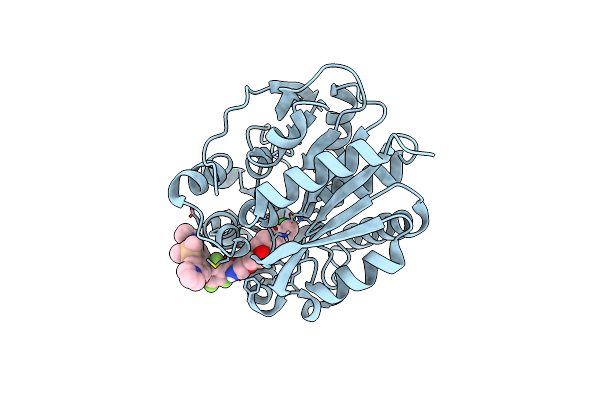
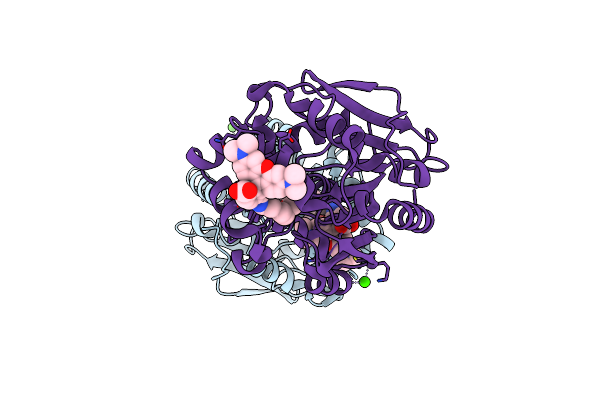
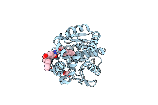
Organism: Homo sapiens
Method: X-RAY DIFFRACTION Resolution:1.45 Å Release Date: 2025-08-20 Classification: HYDROLASE Ligands: A1JLK, ZN, EDO |
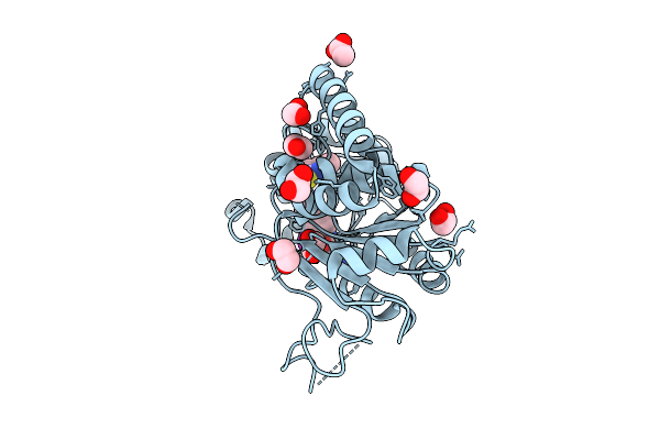 |
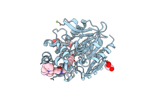
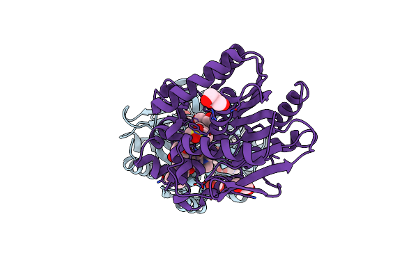
Organism: Homo sapiens
Method: X-RAY DIFFRACTION Resolution:1.45 Å Release Date: 2025-08-20 Classification: HYDROLASE Ligands: ZN, A1JLL, EDO, ACT |
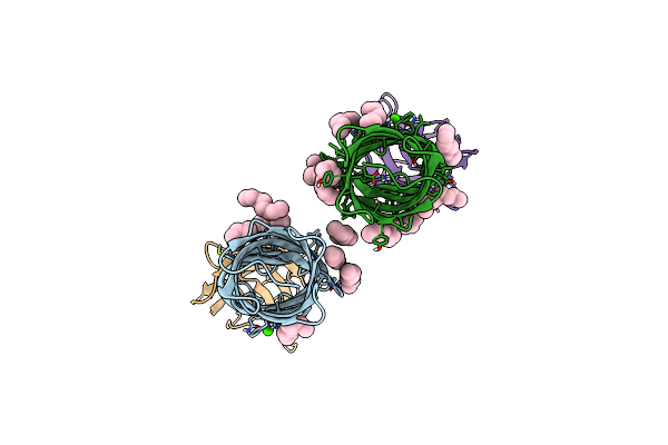 |
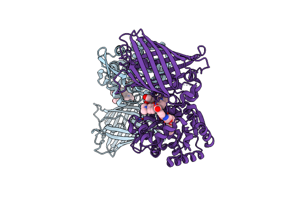
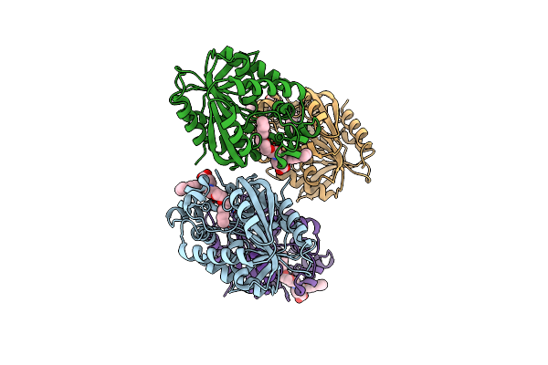
Organism: Escherichia coli k-12, Vicugna pacos
Method: X-RAY DIFFRACTION Resolution:2.27 Å Release Date: 2024-07-17 Classification: MEMBRANE PROTEIN Ligands: OCT, CA |
 |
Organism: Escherichia coli, Vicugna pacos
Method: X-RAY DIFFRACTION Resolution:2.30 Å Release Date: 2024-07-17 Classification: MEMBRANE PROTEIN |
 |
Organism: Rhodococcus sp. (in: high g+c gram-positive bacteria)
Method: X-RAY DIFFRACTION Resolution:1.90 Å Release Date: 2024-06-05 Classification: HYDROLASE Ligands: XSR, CL |
 |
Organism: Saccharomyces cerevisiae
Method: ELECTRON MICROSCOPY Release Date: 2024-01-17 Classification: LIGASE Ligands: PCW, PX6 |
 |
Organism: Saccharomyces cerevisiae
Method: ELECTRON MICROSCOPY Release Date: 2024-01-17 Classification: LIGASE Ligands: PCW, PX6 |
 |
X-Ray Structure Of The Haloalkane Dehalogenase Halotag7 Labeled With A Chloroalkane Cyanine3 Fluorophore Substrate
Organism: Rhodococcus sp.
Method: X-RAY DIFFRACTION Resolution:1.50 Å Release Date: 2023-07-26 Classification: HYDROLASE Ligands: PJI, CL, GOL, MG |
 |
X-Ray Structure Of The Haloalkane Dehalogenase Halotag7 Fusion To The Green Fluorescent Protein Gfp (Chemog1) Labeled With A Chloroalkane Tetramethylrhodamine Fluorophore Substrate
Organism: Rhodococcus sp.
Method: X-RAY DIFFRACTION Resolution:1.80 Å Release Date: 2023-07-26 Classification: HYDROLASE Ligands: OEH, CL, GOL |
 |
X-Ray Structure Of The Interface Optimized Haloalkane Dehalogenase Halotag7 Fusion To The Green Fluorescent Protein Gfp (Chemog5-Tmr) Labeled With A Chloroalkane Tetramethylrhodamine Fluorophore Substrate
Organism: Rhodococcus sp.
Method: X-RAY DIFFRACTION Resolution:2.00 Å Release Date: 2023-07-26 Classification: HYDROLASE Ligands: OEH, CL |
 |
X-Ray Structure Of The Haloalkane Dehalogenase Halotag7 Bound To A Pentyltrifluoromethanesulfonamide Tetramethylrhodamine Ligand (Tmr-T5)
Organism: Rhodococcus sp.
Method: X-RAY DIFFRACTION Resolution:1.70 Å Release Date: 2023-02-22 Classification: HYDROLASE Ligands: IYI, CA |
 |
X-Ray Structure Of The Dead Variant Haloalkane Dehalogenase Halotag7-D106A Bound To A Pentanol Tetramethylrhodamine Ligand (Tmr-Hy5)
Organism: Rhodococcus sp.
Method: X-RAY DIFFRACTION Resolution:1.50 Å Release Date: 2023-02-22 Classification: HYDROLASE Ligands: IYL, CL, GOL |
 |
X-Ray Structure Of The Haloalkane Dehalogenase Halotag7 Bound To A Pentylmethanesulfonamide Tetramethylrhodamine Ligand (Tmr-S5)
Organism: Rhodococcus sp.
Method: X-RAY DIFFRACTION Resolution:1.50 Å Release Date: 2023-02-22 Classification: HYDROLASE Ligands: IYO, GOL |
 |
X-Ray Structure Of The Haloalkane Dehalogenase Halotag7-M175W Labeled With A Chloroalkane-Tetramethylrhodamine Fluorophore Substrate
Organism: Rhodococcus sp.
Method: X-RAY DIFFRACTION Resolution:2.30 Å Release Date: 2021-12-15 Classification: HYDROLASE Ligands: OEH, CL |
 |
X-Ray Structure Of The Haloalkane Dehalogenase Halotag7-Q165W Labeled With A Chloroalkane-Tetramethylrhodamine Fluorophore Substrate
Organism: Rhodococcus sp.
Method: X-RAY DIFFRACTION Resolution:1.40 Å Release Date: 2021-12-15 Classification: HYDROLASE Ligands: OEH, CL, GOL |
 |
X-Ray Structure Of The Haloalkane Dehalogenase Halotag7-P174L Labeled With A Chloroalkane-Tetramethylrhodamine Fluorophore Substrate
Organism: Rhodococcus sp.
Method: X-RAY DIFFRACTION Resolution:1.40 Å Release Date: 2021-08-04 Classification: HYDROLASE Ligands: OEH, CL, GOL |
 |
X-Ray Structure Of The Haloalkane Dehalogenase Halotag7-P174W Labeled With A Chloroalkane-Tetramethylrhodamine Fluorophore Substrate
Organism: Rhodococcus sp.
Method: X-RAY DIFFRACTION Resolution:1.40 Å Release Date: 2021-08-04 Classification: HYDROLASE Ligands: OEH, CL, GOL, CA |
 |
X-Ray Structure Of The Haloalkane Dehalogenase Halotag7-Q165H Labeled With A Chloroalkane-Tetramethylrhodamine Fluorophore Substrate
Organism: Rhodococcus sp.
Method: X-RAY DIFFRACTION Resolution:1.60 Å Release Date: 2021-08-04 Classification: HYDROLASE Ligands: OEH, CL, GOL, PEG |
 |
X-Ray Structure Of The Haloalkane Dehalogenase Halotag7-Q165H-P174L Labeled With A Chloroalkane-Tetramethylrhodamine Fluorophore Substrate
Organism: Rhodococcus sp.
Method: X-RAY DIFFRACTION Resolution:1.40 Å Release Date: 2021-08-04 Classification: HYDROLASE Ligands: OEH, CL, GOL |