
Search Count: 6
 |
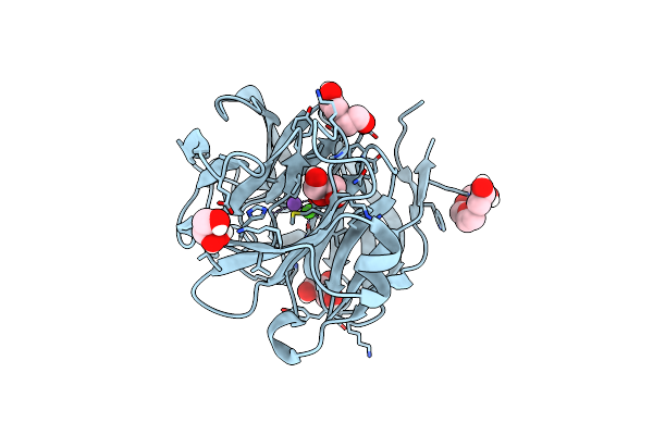
Organism: Escherichia coli o157:h7
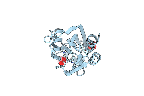
Method: X-RAY DIFFRACTION Resolution:1.94 Å Release Date: 2017-06-21 Classification: HYDROLASE Ligands: SO4, GOL |
 |
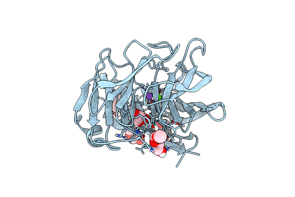
Organism: Escherichia coli o157:h7
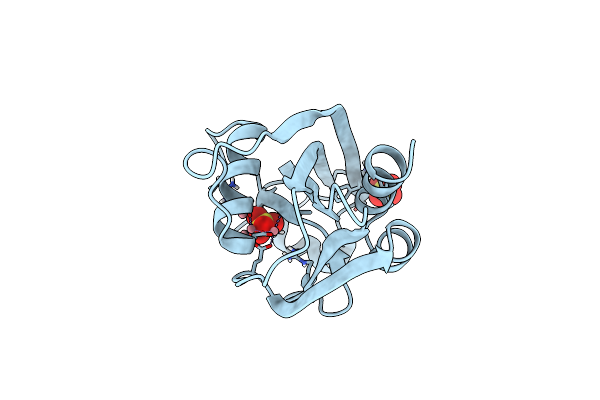
Method: X-RAY DIFFRACTION Resolution:1.79 Å Release Date: 2017-06-21 Classification: HYDROLASE Ligands: SO4, CO |
 |
Organism: Escherichia coli o157:h7
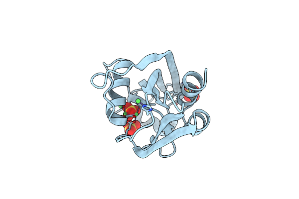
Method: X-RAY DIFFRACTION Resolution:2.00 Å Release Date: 2017-06-21 Classification: HYDROLASE Ligands: SO4, NI |
 |
Organism: Homo sapiens
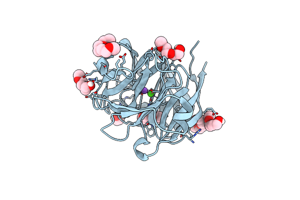
Method: X-RAY DIFFRACTION Resolution:2.15 Å Release Date: 2015-04-01 Classification: PROTEIN BINDING Ligands: CA, P6G, NA |
 |
Crystal Structure Of The E396D Snp Variant Of The Myocilin Olfactomedin Domain
Organism: Homo sapiens
Method: X-RAY DIFFRACTION Resolution:1.90 Å Release Date: 2015-04-01 Classification: PROTEIN BINDING Ligands: CA, GOL, PG4, P6G, NA |
 |
Crystal Structure Of The Selenomthionine Incorporated Myocilin Olfactomedin Domain E396D Variant.
Organism: Homo sapiens
Method: X-RAY DIFFRACTION Resolution:2.09 Å Release Date: 2015-04-01 Classification: PROTEIN BINDING Ligands: CA, P6G, GOL, NA |