
Search Count: 19
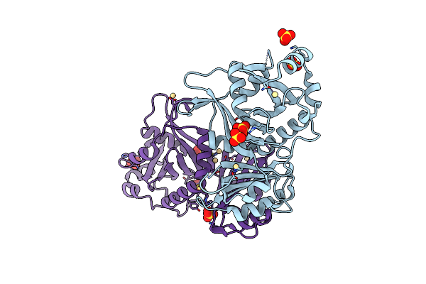 |
Organism: Lambdavirus lambda
Method: X-RAY DIFFRACTION Resolution:2.05 Å Release Date: 2024-04-17 Classification: DNA BINDING PROTEIN Ligands: SO4, CD |
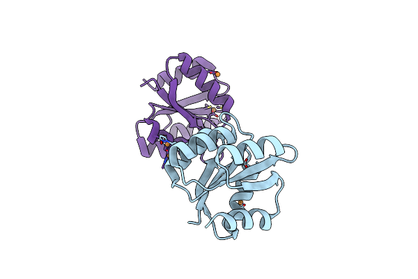 |
Organism: Thermotoga maritima msb8
Method: X-RAY DIFFRACTION Resolution:2.70 Å Release Date: 2018-10-17 Classification: SIGNALING PROTEIN Ligands: CU |
 |
Organism: Human herpesvirus 1
Method: X-RAY DIFFRACTION Resolution:3.60 Å Release Date: 2018-05-16 Classification: VIRAL PROTEIN Ligands: NAG |
 |
Organism: Human herpesvirus 1
Method: X-RAY DIFFRACTION Resolution:4.10 Å Release Date: 2018-05-16 Classification: VIRAL PROTEIN Ligands: NAG |
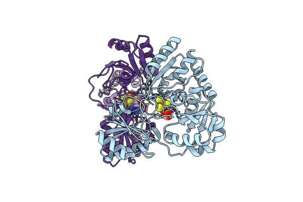 |
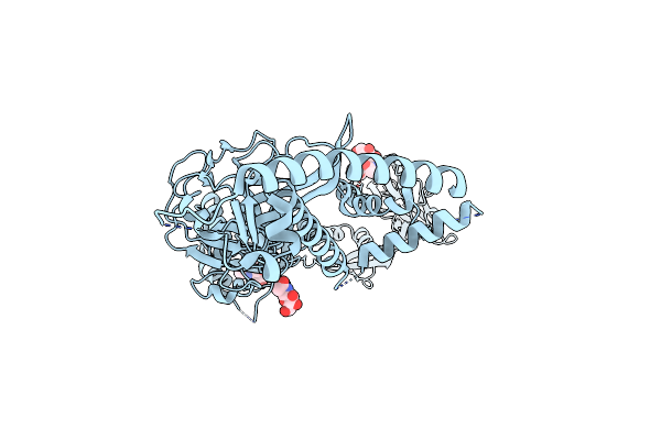
Crystal Structure Of Pyrococcus Horikoshii Dph2 With 4Fe-4S Cluster And Mta
Organism: Pyrococcus horikoshii (strain atcc 700860 / dsm 12428 / jcm 9974 / nbrc 100139 / ot-3)
Method: X-RAY DIFFRACTION Resolution:2.35 Å Release Date: 2018-04-11 Classification: BIOSYNTHETIC PROTEIN Ligands: MTA, SO4, SF4 |
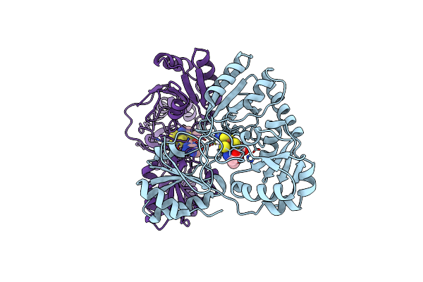 |
Crystal Structure Of Pyrococcus Horikoshii Dph2 With 4Fe-4S Cluster And Sam
Organism: Pyrococcus horikoshii (strain atcc 700860 / dsm 12428 / jcm 9974 / nbrc 100139 / ot-3)
Method: X-RAY DIFFRACTION Resolution:2.30 Å Release Date: 2018-04-11 Classification: BIOSYNTHETIC PROTEIN Ligands: SAM, SF4 |
 |
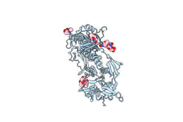
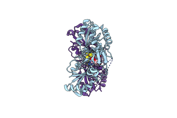
Crystal Structure Of Candidatus Methanoperedens Nitroreducens Dph2 With 4Fe-4S Cluster And Sam/Cleaved Sam
Organism: Candidatus methanoperedens nitroreducens
Method: X-RAY DIFFRACTION Resolution:2.25 Å Release Date: 2018-04-11 Classification: BIOSYNTHETIC PROTEIN Ligands: SF4, SAM, ABA, MTA |
 |
Crystal Structure Of Candidatus Methanoperedens Nitroreducens Dph2 With 4Fe-4S Cluster And Sam
Organism: Candidatus methanoperedens nitroreducens
Method: X-RAY DIFFRACTION Resolution:2.08 Å Release Date: 2018-04-11 Classification: BIOSYNTHETIC PROTEIN Ligands: SF4, SAM |
 |
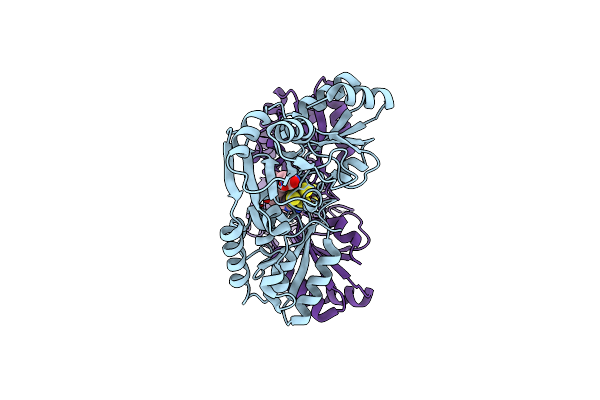
Crystal Structure Of Candidatus Methanoperedens Nitroreducens Dph2 With 4Fe-4S Cluster And Sah
Organism: Candidatus methanoperedens nitroreducens
Method: X-RAY DIFFRACTION Resolution:1.66 Å Release Date: 2018-04-11 Classification: BIOSYNTHETIC PROTEIN Ligands: SF4, SAH |
 |
Organism: Saccharomyces cerevisiae (strain atcc 204508 / s288c)
Method: X-RAY DIFFRACTION Resolution:2.40 Å Release Date: 2015-12-30 Classification: PROTEIN TRANSPORT |
 |
Organism: Saccharomyces cerevisiae (strain atcc 204508 / s288c)
Method: X-RAY DIFFRACTION Resolution:1.60 Å Release Date: 2015-12-30 Classification: PROTEIN TRANSPORT |
 |
Crystal Structure Of A Binary Complex Of Flim-Flig Middle Domains From T.Maritima
Organism: Thermotoga maritima
Method: X-RAY DIFFRACTION Resolution:4.32 Å Release Date: 2015-06-10 Classification: PROTEIN BINDING |
 |
Organism: Pyrococcus horikoshii ot3
Method: X-RAY DIFFRACTION Resolution:4.21 Å Release Date: 2015-02-04 Classification: TRANSPORT PROTEIN Ligands: ASP, NA |
 |
Organism: Homo sapiens
Method: X-RAY DIFFRACTION Resolution:2.38 Å Release Date: 2014-10-15 Classification: OXIDOREDUCTASE Ligands: CU, ZN |
 |
Organism: Pseudomonas aeruginosa
Method: X-RAY DIFFRACTION Resolution:1.95 Å Release Date: 2013-02-27 Classification: SIGNALING PROTEIN Ligands: SO4, NA, CL, MG |
 |
Organism: Pseudomonas aeruginosa
Method: X-RAY DIFFRACTION Resolution:2.88 Å Release Date: 2013-02-27 Classification: SIGNALING PROTEIN Ligands: CL |
 |
Crystal Structure Of The 5-Coordinate Ferric Heme-Binding Pas Domain Of Aer2 From P. Aeruginosa
Organism: Pseudomonas aeruginosa
Method: X-RAY DIFFRACTION Resolution:2.30 Å Release Date: 2013-01-09 Classification: SIGNALING PROTEIN Ligands: HEM, CL, GOL |
 |
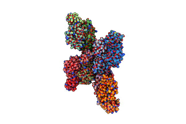
Complex Between Bacterial Chemotaxis Histidine Kinase Chea Domains P4 And P5 And Receptor-Adaptor Protein Chew
Organism: Thermotoga maritima
Method: X-RAY DIFFRACTION Resolution:3.50 Å Release Date: 2006-04-18 Classification: TRANSFERASE/CHEMOTAXIS Ligands: ANP |
 |
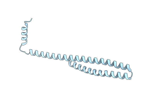
Crystal Structure Of The Cytoplasmic Domain Of A Bacterial Chemoreceptor From Thermotoga Maritima
Organism: Thermotoga maritima
Method: X-RAY DIFFRACTION Resolution:2.50 Å Release Date: 2006-04-18 Classification: CHEMOTAXIS Ligands: PB |