
Search Count: 17
 |
Organism: Lutzomyia longipalpis, Homo sapiens
Method: ELECTRON MICROSCOPY Release Date: 2023-08-30 Classification: IMMUNE SYSTEM Ligands: MG, NAG |
 |
Organism: Lutzomyia longipalpis, Homo sapiens
Method: ELECTRON MICROSCOPY Release Date: 2023-08-09 Classification: IMMUNE SYSTEM Ligands: NAG |
 |
Organism: Lutzomyia longipalpis
Method: X-RAY DIFFRACTION Resolution:2.31 Å Release Date: 2023-08-09 Classification: BLOOD CLOTTING Ligands: GOL, NAG, BR |
 |
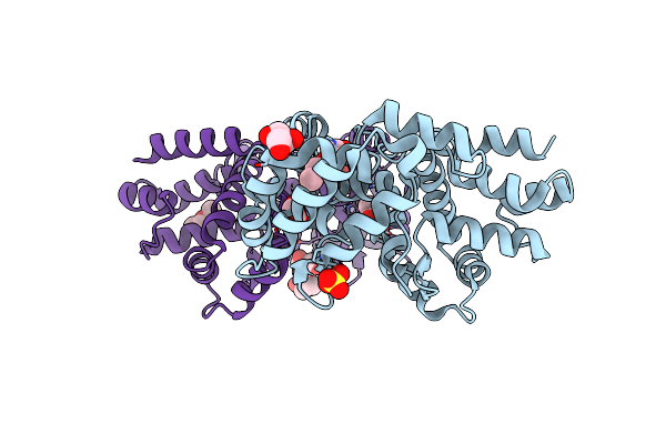
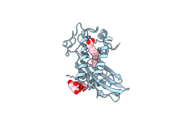
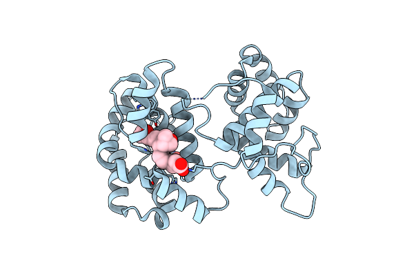
Long Form D7 Protein From Anopheles Darlingi With U46619 And Serotonin Bound
Organism: Anopheles darlingi
Method: X-RAY DIFFRACTION Resolution:2.51 Å Release Date: 2022-06-01 Classification: BLOOD CLOTTING Ligands: SRO, PUC |
 |
Organism: Aedes aegypti
Method: X-RAY DIFFRACTION Resolution:1.19 Å Release Date: 2022-05-25 Classification: BLOOD CLOTTING |
 |
Organism: Culex quinquefasciatus
Method: X-RAY DIFFRACTION Resolution:1.98 Å Release Date: 2022-05-25 Classification: BLOOD CLOTTING Ligands: SO4 |
 |
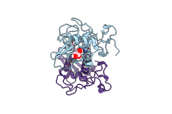
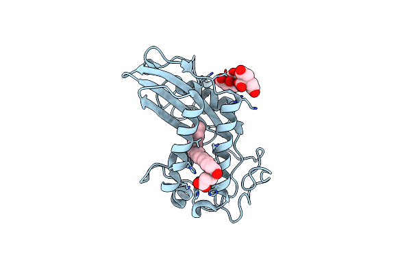
Crystal Structure Of The Anopheles Darlingi Ad-118 Long Form D7 Salivary Protein
Organism: Anopheles darlingi
Method: X-RAY DIFFRACTION Resolution:2.40 Å Release Date: 2022-05-25 Classification: BLOOD CLOTTING Ligands: ETX, SO4, PG0, GOL, PEG, EDO |
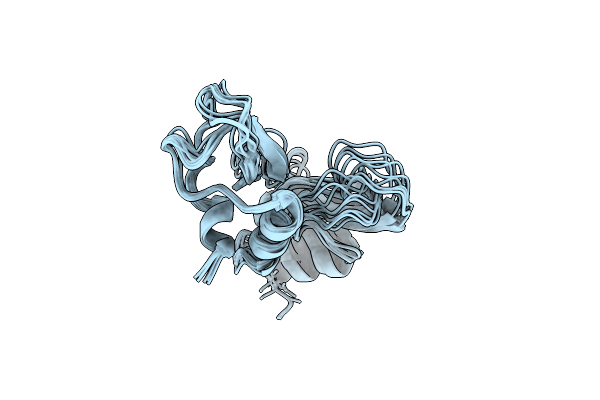 |
Nmr Structure Determination Of Ixolaris And Factor X Interaction Reveals A Noncanonical Mechanism Of Kunitz Inhibition
Organism: Ixodes scapularis
Method: SOLUTION NMR Release Date: 2019-06-12 Classification: BLOOD CLOTTING |
 |
Organism: Ancylostoma caninum
Method: X-RAY DIFFRACTION Resolution:1.60 Å Release Date: 2015-04-22 Classification: BLOOD CLOTTING Ligands: GOL |
 |
Organism: Phlebotomus duboscqi
Method: X-RAY DIFFRACTION Resolution:2.60 Å Release Date: 2013-10-16 Classification: PROTEIN BINDING Ligands: SO4 |
 |
Organism: Tabanus yao
Method: X-RAY DIFFRACTION Resolution:1.57 Å Release Date: 2012-02-15 Classification: PROTEIN BINDING Ligands: PLM, PR, CIT |
 |
Organism: Tabanus yao
Method: X-RAY DIFFRACTION Resolution:1.65 Å Release Date: 2012-02-15 Classification: PROTEIN BINDING Ligands: PR, CIT, PLM |
 |
Organism: Tabanus yao
Method: X-RAY DIFFRACTION Resolution:2.50 Å Release Date: 2012-02-15 Classification: PROTEIN BINDING Ligands: PR, CIT, EAH |
 |
Organism: Anopheles stephensi
Method: X-RAY DIFFRACTION Resolution:1.76 Å Release Date: 2010-12-29 Classification: TRANSPORT PROTEIN Ligands: GOL, SO4 |
 |
Organism: Anopheles stephensi
Method: X-RAY DIFFRACTION Resolution:1.43 Å Release Date: 2010-12-29 Classification: TRANSPORT PROTEIN Ligands: EAH, GOL, SO4 |
 |
Organism: Anopheles stephensi
Method: X-RAY DIFFRACTION Resolution:1.45 Å Release Date: 2010-12-29 Classification: TRANSPORT PROTEIN Ligands: U46, SO4 |
 |
Organism: Ixodes ricinus
Method: X-RAY DIFFRACTION Resolution:1.80 Å Release Date: 2010-10-27 Classification: HYDROLASE INHIBITOR Ligands: 1PE |