
Search Count: 69
 |
Organism: Gallus gallus
Method: X-RAY DIFFRACTION Resolution:0.96 Å Release Date: 2024-11-06 Classification: HYDROLASE Ligands: NO3, ACT |
 |
Organism: Gallus gallus
Method: X-RAY DIFFRACTION Resolution:1.07 Å Release Date: 2024-11-06 Classification: HYDROLASE Ligands: NO3, ACT |
 |
Neutron Structure Of Hydrogenated Hen Egg-White Lysozyme At Room Temperature
Organism: Gallus gallus
Method: NEUTRON DIFFRACTION Resolution:1.10 Å Release Date: 2024-11-06 Classification: HYDROLASE Ligands: NO3, ACT, DOD |
 |
Neutron Structure Of Perdeuterated Hen Egg-White Lysozyme At Room Temperature
Organism: Gallus gallus
Method: NEUTRON DIFFRACTION Resolution:0.91 Å Release Date: 2024-11-06 Classification: HYDROLASE Ligands: NO3, ACT, DOD |
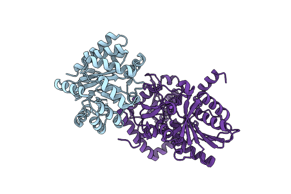 |
Joint X-Ray/Neutron Structure Of Salmonella Typhimurium Tryptophan Synthase Internal Aldimine From Microgravity-Grown Crystal
Organism: Salmonella enterica subsp. enterica serovar typhi, Salmonella enterica subsp. enterica serovar typhimurium
Method: X-RAY DIFFRACTION, NEUTRON DIFFRACTION Resolution:1.80 Å, 2.10 Å Release Date: 2024-02-14 Classification: LYASE |
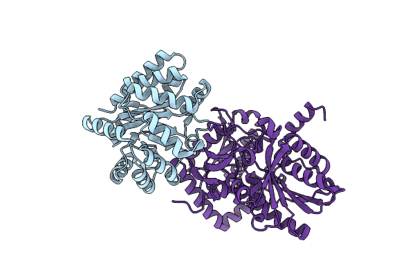 |
X-Ray Crystal Structure Of Salmonella Typhimurium Tryptophan Synthase Internal Aldimine At Ph 5.0
Organism: Salmonella typhimurium (strain lt2 / sgsc1412 / atcc 700720)
Method: X-RAY DIFFRACTION Resolution:2.20 Å Release Date: 2024-02-14 Classification: LYASE |
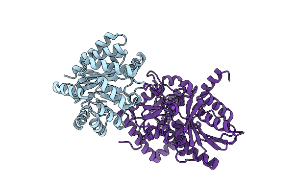 |
X-Ray Crystal Structure Of Salmonella Typhimurium Tryptophan Synthase Internal Aldimine
Organism: Salmonella typhimurium (strain lt2 / sgsc1412 / atcc 700720)
Method: X-RAY DIFFRACTION Resolution:1.60 Å Release Date: 2024-02-14 Classification: LYASE |
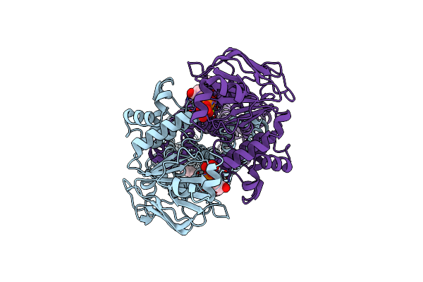 |
Organism: Escherichia coli (strain k12)
Method: ELECTRON MICROSCOPY Release Date: 2022-01-12 Classification: MEMBRANE PROTEIN Ligands: VO4, ADP, MG, POV |
 |
Joint X-Ray/Neutron Room Temperature Structure Of Perdeuterated Lecb Lectin In Complex With Perdeuterated Fucose
Organism: Pseudomonas aeruginosa
Method: X-RAY DIFFRACTION, NEUTRON DIFFRACTION Resolution:1.85 Å, 1.90 Å Release Date: 2022-01-12 Classification: SUGAR BINDING PROTEIN Ligands: CA, FUC, SO4 |
 |
X-Ray Crystal Structure Of Perdeuterated Lecb Lectin In Complex With Perdeuterated Fucose
Organism: Pseudomonas aeruginosa
Method: X-RAY DIFFRACTION Resolution:0.90 Å Release Date: 2022-01-12 Classification: SUGAR BINDING PROTEIN Ligands: FUC, CA, SO4 |
 |
Organism: Gallus gallus
Method: X-RAY DIFFRACTION Resolution:0.89 Å Release Date: 2021-12-22 Classification: HYDROLASE Ligands: NO3, ACT |
 |
X-Ray Crystal Structure Of Aspartate Alpha-Decarboxylase In Complex With D-Serine
Organism: Escherichia coli
Method: X-RAY DIFFRACTION Resolution:1.75 Å Release Date: 2021-10-06 Classification: LYASE Ligands: DSN, EDO, SER |
 |
Organism: Gallus gallus
Method: X-RAY DIFFRACTION Resolution:0.98 Å Release Date: 2021-05-12 Classification: HYDROLASE Ligands: NO3, ACT |
 |
Organism: Gallus gallus
Method: X-RAY DIFFRACTION Resolution:1.00 Å Release Date: 2021-05-12 Classification: HYDROLASE Ligands: NO3, ACT |
 |
Organism: Gallus gallus
Method: X-RAY DIFFRACTION Resolution:1.00 Å Release Date: 2021-05-12 Classification: HYDROLASE Ligands: NO3, ACT |
 |
Joint X-Ray/Neutron Room Temperature Structure Of Perdeuterated Pll Lectin In Complex With Perdeuterated L-Fucose
Organism: Photorhabdus laumondii
Method: X-RAY DIFFRACTION, NEUTRON DIFFRACTION Resolution:1.84 Å, 2.2 Å Release Date: 2021-03-24 Classification: SUGAR BINDING PROTEIN Ligands: FUL, FUC |
 |
Room Temperature X-Ray Structure Of Perdeuterated Pll Lectin In Complex With L-Fucose
Organism: Photorhabdus laumondii
Method: X-RAY DIFFRACTION Resolution:1.55 Å Release Date: 2021-03-17 Classification: SUGAR BINDING PROTEIN Ligands: FUL, FUC |
 |
Organism: Photorhabdus laumondii
Method: X-RAY DIFFRACTION Resolution:1.60 Å Release Date: 2021-03-17 Classification: SUGAR BINDING PROTEIN |
 |
Room Temperature X-Ray Structure Of H/D-Exchanged Pll Lectin In Complex With L-Fucose
Organism: Photorhabdus laumondii
Method: X-RAY DIFFRACTION Resolution:1.60 Å Release Date: 2021-03-17 Classification: SUGAR BINDING PROTEIN Ligands: FUC, FUL |
 |
Organism: Photorhabdus laumondii
Method: X-RAY DIFFRACTION Resolution:1.70 Å Release Date: 2021-03-17 Classification: SUGAR BINDING PROTEIN Ligands: FUL, FUC, GOL |