
Search Count: 41
All
Selected
 |
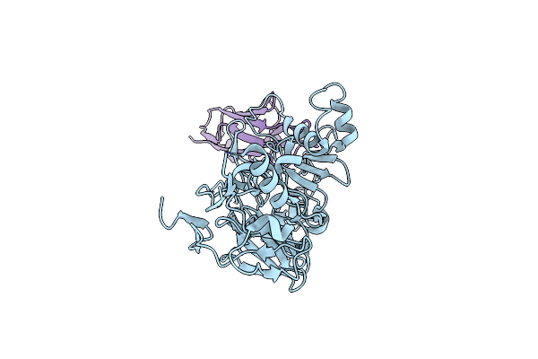
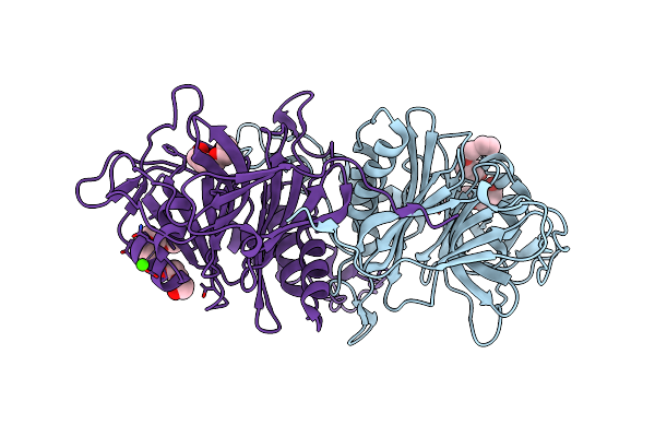
Crystal Structure Of The Plasmodium Falciparum Apical Membrane Antigen (Ama1) In Complex With Single Domain I-Body Wd34
Organism: Plasmodium falciparum 3d7, Homo sapiens
Method: X-RAY DIFFRACTION Resolution:2.60 Å Release Date: 2024-07-03 Classification: PROTEIN BINDING Ligands: MPD, EPE, NA |
 |
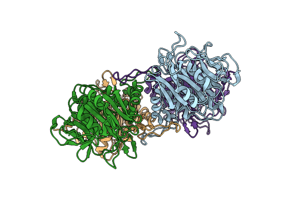
Crystal Structure Of The Plasmodium Vivax Apical Membrane Antigen (Ama1) In Complex With Single Domain I-Body Wd34
Organism: Plasmodium vivax sal-1, Homo sapiens
Method: X-RAY DIFFRACTION Resolution:3.00 Å Release Date: 2024-07-03 Classification: PROTEIN BINDING |
 |
Organism: Lactobacillus acidophilus
Method: X-RAY DIFFRACTION Resolution:2.15 Å Release Date: 2023-01-25 Classification: HYDROLASE |
 |
Organism: Lactobacillus gasseri
Method: X-RAY DIFFRACTION Resolution:2.05 Å Release Date: 2023-01-25 Classification: HYDROLASE Ligands: TAU, K |
 |
Bile Salt Hydrolase A From Lactobacillus Gasseri With Chenodeoxycholate And Taurine Bound
Organism: Lactobacillus gasseri
Method: X-RAY DIFFRACTION Resolution:1.35 Å Release Date: 2023-01-25 Classification: HYDROLASE Ligands: TAU, JN3 |
 |
Organism: Lactobacillus gasseri
Method: X-RAY DIFFRACTION Resolution:1.56 Å Release Date: 2023-01-25 Classification: HYDROLASE Ligands: MG |
 |
Organism: Lactobacillus johnsonii
Method: X-RAY DIFFRACTION Resolution:1.45 Å Release Date: 2023-01-25 Classification: HYDROLASE |
 |
Organism: Lactobacillus ingluviei
Method: X-RAY DIFFRACTION Resolution:1.43 Å Release Date: 2023-01-25 Classification: HYDROLASE Ligands: PG4, PEG, PGE, CA |
 |
Organism: Lactobacillus reuteri
Method: X-RAY DIFFRACTION Resolution:1.66 Å Release Date: 2023-01-25 Classification: HYDROLASE Ligands: SO4 |
 |
Organism: Homo sapiens
Method: X-RAY DIFFRACTION Resolution:2.20 Å Release Date: 2021-10-06 Classification: SIGNALING PROTEIN Ligands: 1S2, ACT, DMS, EDO |
 |
Organism: Homo sapiens
Method: X-RAY DIFFRACTION Resolution:2.28 Å Release Date: 2021-09-22 Classification: TRANSFERASE Ligands: ZN, SAM, YJ1, BME |
 |
Organism: Homo sapiens
Method: X-RAY DIFFRACTION Resolution:1.80 Å Release Date: 2021-09-22 Classification: TRANSFERASE Ligands: ZN, SAM, YHY, EDO, DMS |
 |
Organism: Homo sapiens
Method: X-RAY DIFFRACTION Resolution:2.47 Å Release Date: 2021-09-22 Classification: TRANSFERASE Ligands: ZN, SAM, YHV, EDO |
 |
Macrocyclic Peptides Tdi5575 That Selectively Inhibit The Mycobacterium Tuberculosis Proteasome
Organism: Mycobacterium tuberculosis
Method: X-RAY DIFFRACTION Resolution:2.28 Å Release Date: 2021-04-28 Classification: HYDROLASE/HYDROLASE INHIBITOR Ligands: U5Y, CIT, DMF |
 |
Organism: Mycobacterium tuberculosis (strain atcc 25618 / h37rv)
Method: X-RAY DIFFRACTION Resolution:2.21 Å Release Date: 2021-01-27 Classification: OXIDOREDUCTASE/OXIDOREDUCTASE INHIBITOR Ligands: FAD, WPM, GOL |
 |
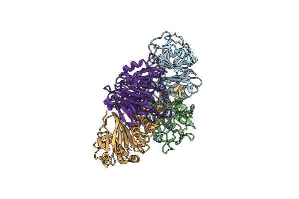
Crystal Structure Of Mycobacterium Tuberculosis Proteasome In Complex With Phenylimidazole-Based Inhibitor A85
Organism: Mycobacterium tuberculosis (strain atcc 25618 / h37rv)
Method: X-RAY DIFFRACTION Resolution:2.60 Å Release Date: 2019-10-09 Classification: HYDROLASE/HYDROLASE INHIBITOR Ligands: DMF, M6M, CIT |
 |
Crystal Structure Of Mycobacterium Tuberculosis Proteasome In Complex With Phenylimidazole-Based Inhibitor A86
Organism: Mycobacterium tuberculosis (strain atcc 25618 / h37rv)
Method: X-RAY DIFFRACTION Resolution:2.65 Å Release Date: 2019-10-09 Classification: HYDROLASE/HYDROLASE INHIBITOR Ligands: DMF, M6Y, CIT |
 |
Crystal Structure Of Mycobacterium Tuberculosis Proteasome In Complex With Phenylimidazole-Based Inhibitor B6
Organism: Mycobacterium tuberculosis (strain atcc 25618 / h37rv)
Method: X-RAY DIFFRACTION Resolution:2.90 Å Release Date: 2019-10-09 Classification: HYDROLASE/HYDROLASE INHIBITOR Ligands: M9G |
 |
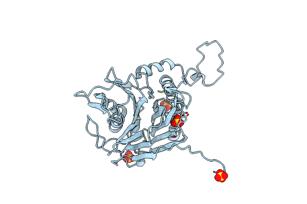
Organism: Homo sapiens
Method: X-RAY DIFFRACTION Resolution:3.01 Å Release Date: 2019-09-25 Classification: CELL ADHESION Ligands: NAG, MN, JUY |
 |
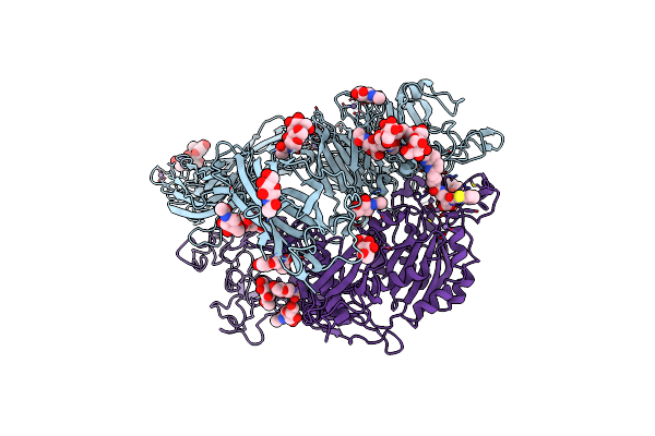
Bacteroides Ovatus Mixed-Linkage Glucan Utilization Locus (Mlgul) Sgbp-A With Cellohexaose
Organism: Bacteroides ovatus (strain atcc 8483 / dsm 1896 / jcm 5824 / nctc 11153)
Method: X-RAY DIFFRACTION Resolution:2.40 Å Release Date: 2019-05-29 Classification: SUGAR BINDING PROTEIN Ligands: MG, ACT, PEG, EDO |