
Search Count: 36
 |
Organism: Bacillus subtilis (strain 168)
Method: X-RAY DIFFRACTION Resolution:2.70 Å Release Date: 2016-01-20 Classification: REPLICATION |
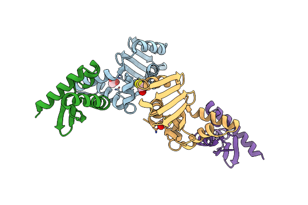 |
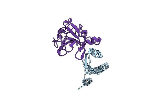
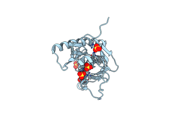
Sporulation Inhibitor Of Dna Replication, Sira, In Complex With Domain I Of Dnaa
Organism: Bacillus subtilis
Method: X-RAY DIFFRACTION Resolution:1.65 Å Release Date: 2014-07-30 Classification: REPLICATION Ligands: BME, ACT |
 |
Organism: Bacillus subtilis
Method: X-RAY DIFFRACTION Resolution:2.26 Å Release Date: 2012-03-14 Classification: SIGNALING PROTEIN |
 |
Organism: Bacillus subtilis
Method: X-RAY DIFFRACTION Resolution:1.90 Å Release Date: 2011-06-15 Classification: TRANSCRIPTION |
 |
Organism: Bacillus subtilis
Method: X-RAY DIFFRACTION Resolution:2.27 Å Release Date: 2011-06-08 Classification: TRANSCRIPTION Ligands: NI |
 |
The Crystal Structure Of Leishmania Major Dutpase In Complex With Substrate Analogue Dupnpp
Organism: Leishmania major
Method: X-RAY DIFFRACTION Resolution:1.86 Å Release Date: 2011-03-16 Classification: HYDROLASE Ligands: DUP, CA |
 |
Organism: Leishmania major
Method: X-RAY DIFFRACTION Resolution:2.40 Å Release Date: 2011-03-16 Classification: HYDROLASE Ligands: UMP, SO4, MG |
 |
Organism: Leishmania major
Method: X-RAY DIFFRACTION Resolution:2.28 Å Release Date: 2011-03-16 Classification: HYDROLASE Ligands: DUR, SO4 |
 |
Organism: Bacillus subtilis
Method: X-RAY DIFFRACTION Resolution:1.74 Å Release Date: 2011-02-23 Classification: HYDROLASE |
 |
Organism: Bacillus subtilis
Method: X-RAY DIFFRACTION Resolution:2.55 Å Release Date: 2011-02-23 Classification: HYDROLASE Ligands: DUP, MG |
 |
Organism: Bacillus subtilis
Method: X-RAY DIFFRACTION Resolution:1.89 Å Release Date: 2011-02-23 Classification: HYDROLASE Ligands: DUD |
 |
Organism: Plasmodium falciparum
Method: X-RAY DIFFRACTION Resolution:1.89 Å Release Date: 2010-04-21 Classification: TRANSFERASE Ligands: TMP, ADP, NA, GOL |
 |
Organism: Plasmodium falciparum
Method: X-RAY DIFFRACTION Resolution:2.40 Å Release Date: 2010-04-21 Classification: TRANSFERASE Ligands: DGP, ADP, NA, GOL |
 |
Organism: Plasmodium falciparum
Method: X-RAY DIFFRACTION Resolution:2.70 Å Release Date: 2010-04-21 Classification: TRANSFERASE Ligands: T5A, NA |
 |
Organism: Plasmodium falciparum
Method: X-RAY DIFFRACTION Resolution:2.99 Å Release Date: 2010-04-21 Classification: TRANSFERASE Ligands: ATM, ADP |
 |
Crystal Structure Of Rph, An Exoribonuclease From Bacillus Anthracis At 1.7 A Resolution
Organism: Bacillus anthracis
Method: X-RAY DIFFRACTION Resolution:1.70 Å Release Date: 2009-02-10 Classification: TRANSFERASE Ligands: SO4 |
 |
Crystal Structure Of Ferrochelatase Hemh-1 From Bacillus Anthracis, Str. Ames
Organism: Bacillus anthracis
Method: X-RAY DIFFRACTION Resolution:2.10 Å Release Date: 2007-05-01 Classification: LYASE |
 |
The Crystal Structure Of A Complex Of Leishmania Major Dutpase With Substrate Analogue Dupnhp
Organism: Leishmania major
Method: X-RAY DIFFRACTION Resolution:2.34 Å Release Date: 2007-04-17 Classification: HYDROLASE Ligands: DUN, MG |
 |
The Crystal Structure Of A Complex Of Campylobacter Jejuni Dutpase With Substrate Analogue Dupnhpp
Organism: Campylobacter jejuni
Method: X-RAY DIFFRACTION Release Date: 2007-03-27 Classification: HYDROLASE Ligands: DUP, MG |
 |
Crystal Structure Of Inosine-Uridine Preferring Nucleoside Hydrolase From Bacillus Anthracis At 2.2A Resolution
Organism: Bacillus anthracis
Method: X-RAY DIFFRACTION Resolution:2.20 Å Release Date: 2007-02-27 Classification: HYDROLASE Ligands: CA, RIB |