
Search Count: 18
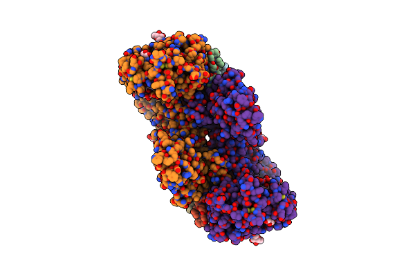 |
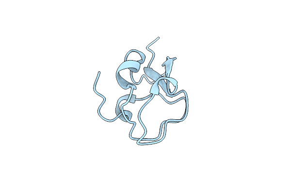
The Insulin Receptor Ectodomain In Complex With A Venom Hybrid Insulin Analog - "Head" Region
Organism: Homo sapiens
Method: ELECTRON MICROSCOPY Release Date: 2022-03-16 Classification: HORMONE,TOXIN Ligands: NAG |
 |
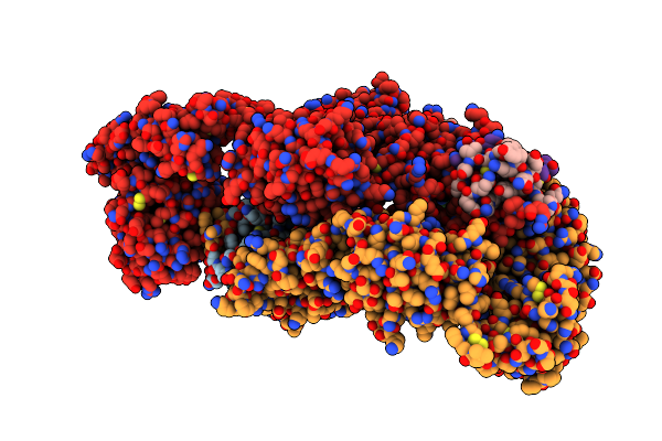
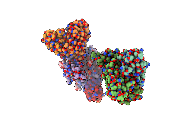
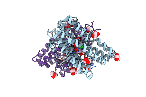
The Insulin Receptor Ectodomain In Complex With Four Venom Hybrid Insulins - Symmetric Conformation
Organism: Homo sapiens
Method: ELECTRON MICROSCOPY Release Date: 2022-03-16 Classification: HORMONE,TOXIN Ligands: NAG |
 |
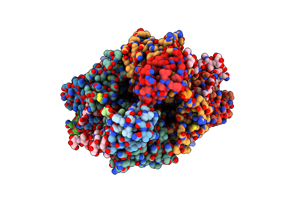
The Insulin Receptor Ectodomain In Complex With Three Venom Hybrid Insulin Molecules - Asymmetric Conformation
Organism: Homo sapiens
Method: ELECTRON MICROSCOPY Release Date: 2022-03-16 Classification: HORMONE,TOXIN |
 |
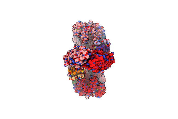
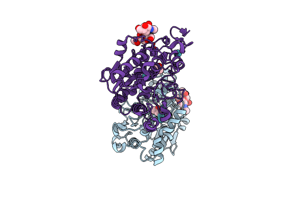
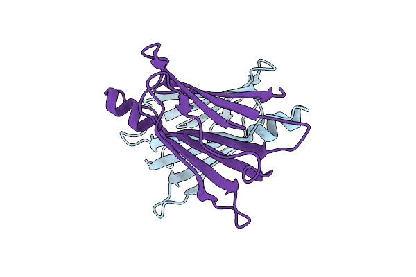
Human Insulin In Complex With The Human Insulin Microreceptor In Turn In Complex With Fv 83-7
Organism: Homo sapiens, Mus musculus
Method: X-RAY DIFFRACTION Resolution:2.90 Å Release Date: 2020-06-03 Classification: HORMONE RECEPTOR/HORMONE/IMMUNE SYSTEM Ligands: NAG |
 |
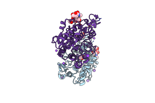
Con-Ins G1 In Complex With The Human Insulin Microreceptor In Turn In Complex With Fv 83-7
Organism: Homo sapiens, Mus musculus, Conus geographus
Method: X-RAY DIFFRACTION Resolution:3.25 Å Release Date: 2020-06-03 Classification: TOXIN Ligands: NAG, SO4 |
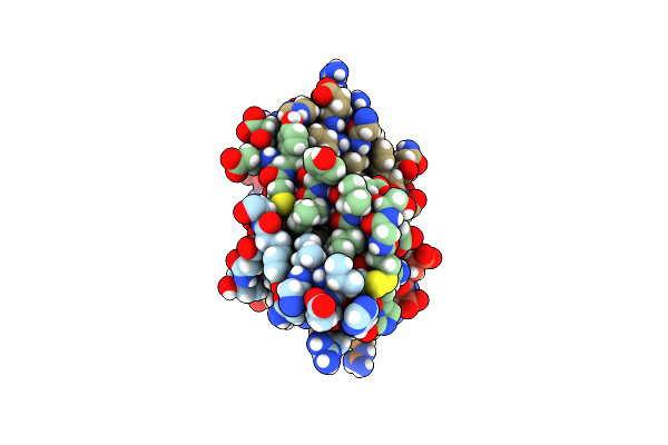 |
Organism: Homo sapiens
Method: X-RAY DIFFRACTION Resolution:1.46 Å Release Date: 2020-06-03 Classification: HORMONE |
 |
Open And Closed Conformations And Protonation States Of Candida Antarctica Lipase B: Xenon Complex
Organism: Pseudozyma antarctica
Method: X-RAY DIFFRACTION Resolution:2.28 Å Release Date: 2015-10-21 Classification: HYDROLASE Ligands: IPA, XE |
 |
Open And Closed Conformations And Protonation States Of Candida Antarctica Lipase B: Atomic Resolution Native
Organism: Pseudozyma antarctica
Method: X-RAY DIFFRACTION Resolution:0.91 Å Release Date: 2015-10-21 Classification: HYDROLASE Ligands: IPA, K, NA |
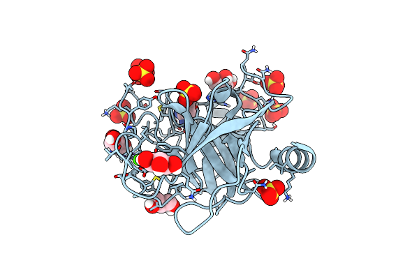 |
Bond Length Analysis Of Asp, Glu And His Residues In Trypsin At 1.2A Resolution
Organism: Bos taurus
Method: X-RAY DIFFRACTION Resolution:1.20 Å Release Date: 2015-05-27 Classification: HYDROLASE Ligands: GOL, SO4, CA, BEN |
 |
Bond Length Analysis Of The Pqqc Y175F Mutant Structure Shows Evidence For Bound Pqq In The Reduced Form
Organism: Klebsiella pneumoniae subsp. pneumoniae
Method: X-RAY DIFFRACTION Resolution:1.44 Å Release Date: 2015-03-04 Classification: OXIDOREDUCTASE Ligands: PQQ, GOL, CL |
 |
Neutron Structure Of Leucine And Valine Methyl Protonated Type Iii Antifreeze
Organism: Zoarces americanus
Method: NEUTRON DIFFRACTION, X-RAY DIFFRACTION Resolution:1.050 Å, 1.850 Å Release Date: 2014-12-24 Classification: ANTIFREEZE PROTEIN Ligands: DOD |
 |
X-Ray Structure Of Human Transthyretin (Ttr) At Room Temperature To 1.9A Resolution
Organism: Homo sapiens
Method: X-RAY DIFFRACTION Resolution:1.85 Å Release Date: 2014-11-12 Classification: TRANSPORT PROTEIN |
 |
Neutron Structure Of Human Transthyretin (Ttr) At Room Temperature To 2.0A Resolution (Laue)
Organism: Homo sapiens
Method: NEUTRON DIFFRACTION, X-RAY DIFFRACTION Resolution:2.0000 Å, 1.9500 Å Release Date: 2014-11-12 Classification: TRANSPORT PROTEIN Ligands: DOD |
 |
Neutron Structure Of Human Transthyretin (Ttr) At Room Temperature To 2.3A Resolution (Monochromatic)
Organism: Homo sapiens
Method: NEUTRON DIFFRACTION, X-RAY DIFFRACTION Resolution:2.3000 Å, 1.9500 Å Release Date: 2014-11-12 Classification: TRANSPORT PROTEIN Ligands: DOD |
 |
Bond Length Analysis Of Asp, Glu And His Residues In Subtilisin Carlsberg At 1.26A Resolution
Organism: Bacillus licheniformis
Method: X-RAY DIFFRACTION Resolution:1.26 Å Release Date: 2012-06-27 Classification: HYDROLASE Ligands: GOL, CA, NA |
 |
Neutron Structure Of Type-Iii Antifreeze Protein Allows The Reconstruction Of Afp-Ice Interface
Organism: Macrozoarces americanus
Method: NEUTRON DIFFRACTION Resolution:1.85 Å Release Date: 2011-06-22 Classification: ANTIFREEZE PROTEIN Ligands: DOD |
 |
Crystal Structure Of The Coenzyme Pqq Synthesis Protein (Pqqb) From Pseudomonas Putida
Organism: Pseudomonas putida
Method: X-RAY DIFFRACTION Resolution:2.20 Å Release Date: 2010-09-22 Classification: BIOSYNTHETIC PROTEIN Ligands: ZN, CL |
 |
Crystal Structure Of The C1 Domain Of Cardiac Isoform Of Myosin Binding Protein-C At 1.3A
Organism: Homo sapiens
Method: X-RAY DIFFRACTION Resolution:1.30 Å Release Date: 2008-07-01 Classification: CONTRACTILE PROTEIN |