
Search Count: 24
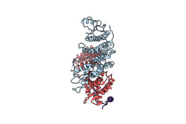 |
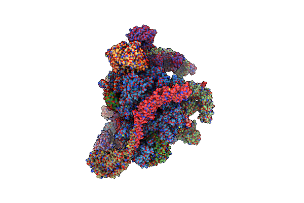
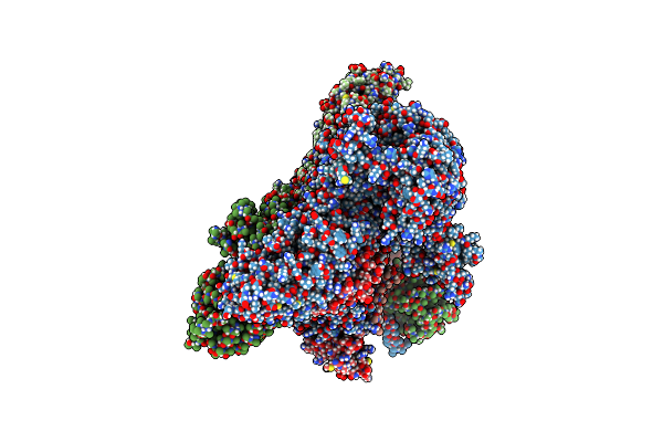
Organism: Homo sapiens
Method: ELECTRON MICROSCOPY Release Date: 2025-06-11 Classification: GENE REGULATION |
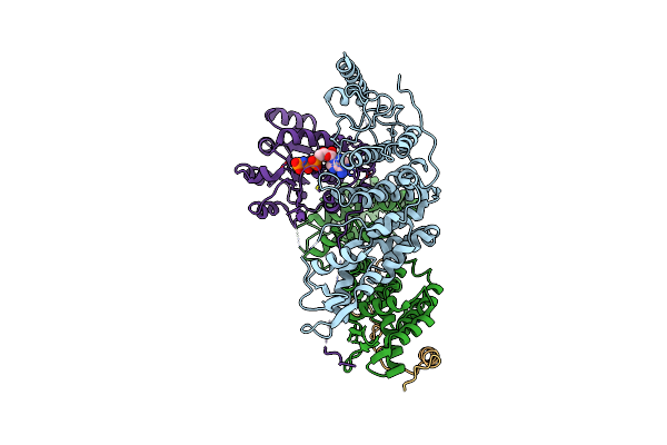 |
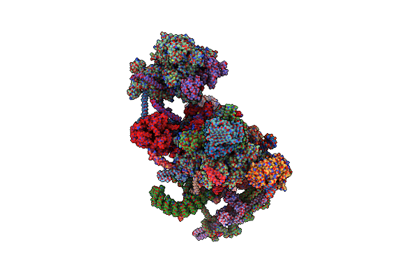
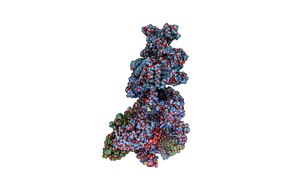
Organism: Homo sapiens
Method: ELECTRON MICROSCOPY Release Date: 2025-06-11 Classification: GENE REGULATION Ligands: ANP |
 |
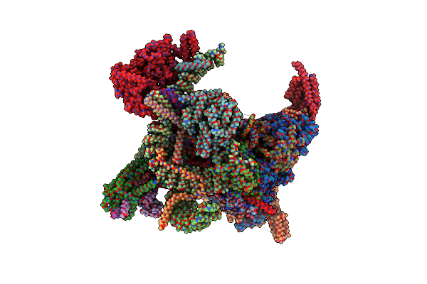
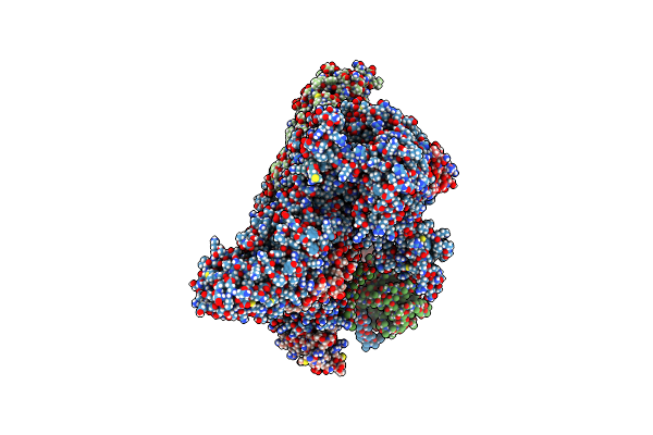
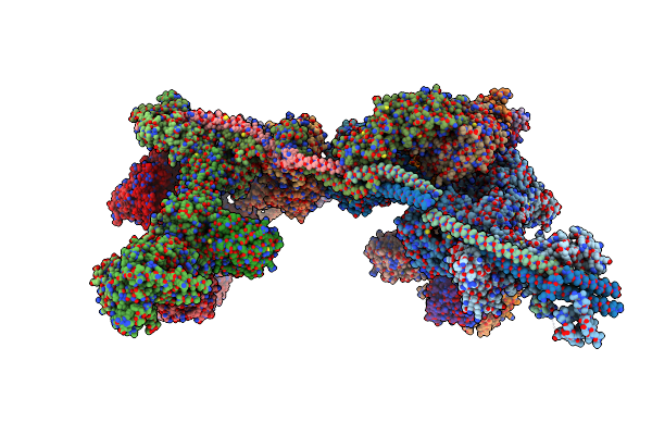
Organism: Homo sapiens
Method: ELECTRON MICROSCOPY Release Date: 2025-06-11 Classification: GENE REGULATION |
 |
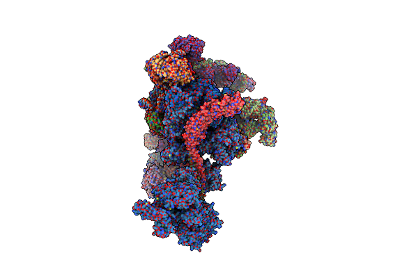
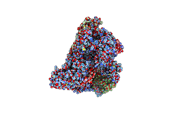
Organism: Homo sapiens
Method: ELECTRON MICROSCOPY Release Date: 2024-09-18 Classification: SPLICING Ligands: GTP, ZN, IHP |
 |
Structure Of The C. Elegans Intron Lariat Spliceosome Primed For Disassembly (Ils')
Organism: Caenorhabditis elegans
Method: ELECTRON MICROSCOPY Release Date: 2024-08-07 Classification: SPLICING Ligands: MG, IHP, GTP, ZN |
 |
Structure Of The C. Elegans Intron Lariat Spliceosome Double-Primed For Disassembly (Ils'')
Organism: Caenorhabditis elegans
Method: ELECTRON MICROSCOPY Release Date: 2024-08-07 Classification: SPLICING Ligands: MG, IHP, GTP, ZN |
 |
Organism: Homo sapiens, Human adenovirus 2
Method: ELECTRON MICROSCOPY Release Date: 2024-07-10 Classification: SPLICING Ligands: MG, K, ATP, GTP, ZN, IHP |
 |
Structure Of The Recycling U5 Snrnp Bound To Chaperones Cd2Bp2 And Tssc4 (State 2)
Organism: Homo sapiens
Method: ELECTRON MICROSCOPY Release Date: 2024-04-17 Classification: SPLICING Ligands: MG, GTP |
 |
Structure Of The Recycling U5 Snrnp Bound To Chaperones Cd2Bp2 And Tssc4 (State 1)
Organism: Homo sapiens
Method: ELECTRON MICROSCOPY Release Date: 2024-04-17 Classification: SPLICING Ligands: MG, GTP |
 |
Organism: Homo sapiens
Method: ELECTRON MICROSCOPY Release Date: 2024-04-17 Classification: SPLICING Ligands: MG, GTP |
 |
Organism: Homo sapiens
Method: ELECTRON MICROSCOPY Release Date: 2024-04-17 Classification: SPLICING Ligands: MG, GTP |
 |
Organism: Homo sapiens
Method: ELECTRON MICROSCOPY Release Date: 2023-05-17 Classification: GENE REGULATION |
 |
Organism: Homo sapiens
Method: ELECTRON MICROSCOPY Release Date: 2023-05-03 Classification: GENE REGULATION |
 |
Organism: Homo sapiens
Method: ELECTRON MICROSCOPY Release Date: 2023-04-12 Classification: GENE REGULATION Ligands: MG, ANP |
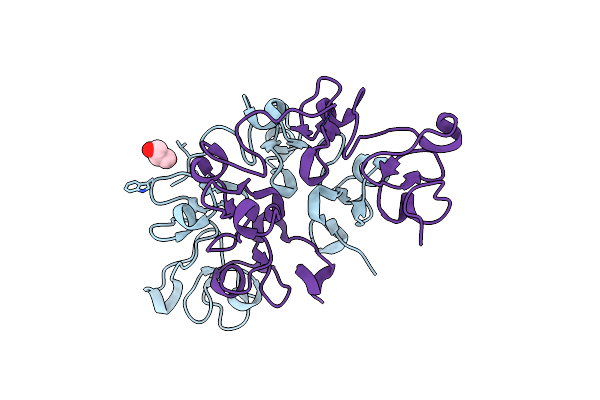 |
Organism: Arundo donax
Method: X-RAY DIFFRACTION Resolution:1.70 Å Release Date: 2021-07-14 Classification: SUGAR BINDING PROTEIN Ligands: GOL |
 |
Three Dimensional Structure Of The Giant Reed (Arundodonax) Lectin (Adl) Complex With N-Acetyl Glucosamine
Organism: Arundo donax
Method: X-RAY DIFFRACTION Resolution:1.75 Å Release Date: 2021-07-14 Classification: SUGAR BINDING PROTEIN Ligands: NAG, GOL |
 |
Three Dimensional Structure Of The Giant Reed (Arundodonax) Lectin (Adl) Complex With N-Acetyl Lactosamine
Organism: Arundo donax
Method: X-RAY DIFFRACTION Resolution:1.50 Å Release Date: 2021-07-14 Classification: SUGAR BINDING PROTEIN Ligands: NAG, GOL |
 |
Three Dimensional Structure Of The Giant Reed (Arundodonax) Lectin (Adl) Complex With Sialic Acid
Organism: Arundo donax
Method: X-RAY DIFFRACTION Resolution:1.60 Å Release Date: 2021-07-14 Classification: SUGAR BINDING PROTEIN Ligands: SIA, GOL |
 |
Three Dimensional Structure Of The Giant Reed (Arundodonax) Lectin (Adl) Complex With N,N'-Diacetylchitobiose; 30 Seconds Soaking
Organism: Arundo donax
Method: X-RAY DIFFRACTION Resolution:1.50 Å Release Date: 2021-07-14 Classification: SUGAR BINDING PROTEIN Ligands: NAG, GOL |
 |
Three Dimensional Structure Of The Giant Reed (Arundodonax) Lectin (Adl) Complex With N,N'-Diacetylchitobiose; 60 Seconds Soaking
Organism: Arundo donax
Method: X-RAY DIFFRACTION Resolution:1.80 Å Release Date: 2021-07-14 Classification: SUGAR BINDING PROTEIN Ligands: NAG, GOL |