
Search Count: 48
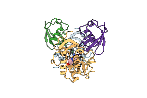 |
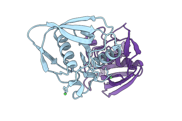
X-Ray Structure Of Turnip Yellow Mosaic Virus Pro/Dub In Complex With Ubiquitin
Organism: Turnip yellow mosaic virus, Homo sapiens
Method: X-RAY DIFFRACTION Resolution:3.66 Å Release Date: 2020-08-05 Classification: VIRAL PROTEIN Ligands: GVE |
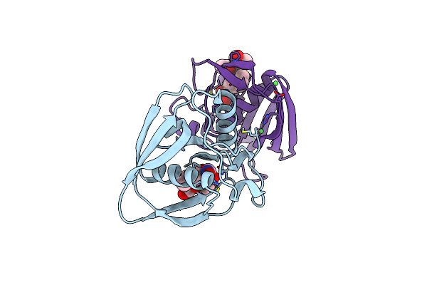 |
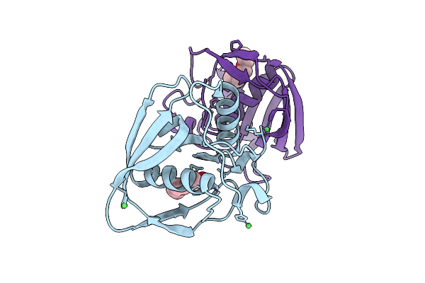
Crystal Structure Of Pdf From The Vibrio Parahaemolyticus Bacteriophage Vp16T In Complex With Actinonin - Crystal Form Ii
Organism: Vibrio phage vp16t
Method: X-RAY DIFFRACTION Resolution:1.40 Å Release Date: 2017-11-29 Classification: HYDROLASE Ligands: BB2, ZN, NI |
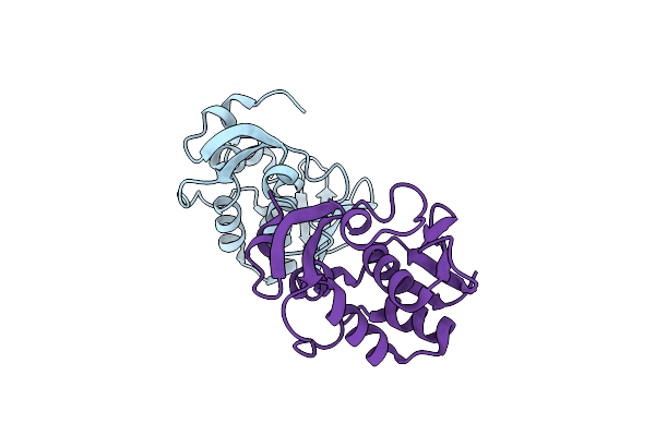 |
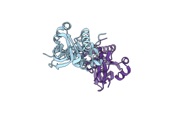
Organism: Turnip yellow mosaic virus
Method: X-RAY DIFFRACTION Resolution:1.65 Å Release Date: 2017-10-25 Classification: HYDROLASE |
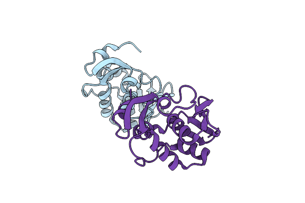 |
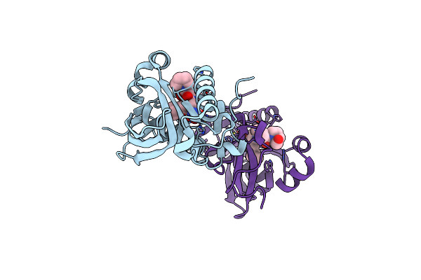
Organism: Turnip yellow mosaic virus
Method: X-RAY DIFFRACTION Resolution:1.65 Å Release Date: 2017-10-25 Classification: HYDROLASE |
 |
Crystal Structure Of Pdf From The Vibrio Parahaemolyticus Bacteriophage Vp16T - Crystal Form I
Organism: Vibrio phage vp16t
Method: X-RAY DIFFRACTION Resolution:1.70 Å Release Date: 2017-09-20 Classification: HYDROLASE Ligands: ZN, NI |
 |
Crystal Structure Of Pdf From The Vibrio Parahaemolyticus Bacteriophage Vp16T - Crystal Form Ii
Organism: Vibrio phage vp16t
Method: X-RAY DIFFRACTION Resolution:1.50 Å Release Date: 2017-09-20 Classification: HYDROLASE Ligands: PGE, ZN, NI |
 |
Crystal Structure Of Type 2 Pdf From Streptococcus Agalactiae, Crystallized In Imidazole Buffer
Organism: Streptococcus agalactiae
Method: X-RAY DIFFRACTION Resolution:2.00 Å Release Date: 2016-11-30 Classification: HYDROLASE Ligands: IMD, ZN |
 |
Crystal Structure Of Type 2 Pdf From Streptococcus Agalactiae, Crystallized In Cacodylate Buffer
Organism: Streptococcus agalactiae
Method: X-RAY DIFFRACTION Resolution:2.80 Å Release Date: 2016-11-30 Classification: HYDROLASE Ligands: NI |
 |
Crystal Structure Of Type 2 Pdf From Streptococcus Agalactiae In Complex With Tripeptide Met-Ala-Ser
Organism: Streptococcus agalactiae, Escherichia coli
Method: X-RAY DIFFRACTION Resolution:1.70 Å Release Date: 2016-11-30 Classification: HYDROLASE Ligands: ACT, ZN |
 |
Crystal Structure Of Type 2 Pdf From Streptococcus Agalactiae In Complex With Tripeptide Met-Ala-Arg
Organism: Streptococcus agalactiae, Escherichia coli
Method: X-RAY DIFFRACTION Resolution:1.60 Å Release Date: 2016-11-30 Classification: HYDROLASE Ligands: ACT, NI |
 |
Crystal Structure Of Type 2 Pdf From Streptococcus Agalactiae In Complex With Actinonin
Organism: Streptococcus agalactiae
Method: X-RAY DIFFRACTION Resolution:2.00 Å Release Date: 2016-11-30 Classification: HYDROLASE Ligands: BB2, ACT, ZN |
 |
Crystal Structure Of Type 2 Pdf From Streptococcus Agalactiae In Complex With Inhibitor At002
Organism: Streptococcus agalactiae
Method: X-RAY DIFFRACTION Resolution:2.00 Å Release Date: 2016-11-30 Classification: HYDROLASE Ligands: SF7, ACT, IMD, ZN |
 |
Crystal Structure Of Type 2 Pdf From Streptococcus Agalactiae In Complex With Inhibitor At018
Organism: Streptococcus agalactiae
Method: X-RAY DIFFRACTION Resolution:1.60 Å Release Date: 2016-11-30 Classification: HYDROLASE Ligands: SF5, ACT, IMD, ZN |
 |
Crystal Structure Of Type 2 Pdf From Streptococcus Agalactiae In Complex With Inhibitor At019
Organism: Streptococcus agalactiae
Method: X-RAY DIFFRACTION Resolution:2.40 Å Release Date: 2016-11-30 Classification: HYDROLASE Ligands: 6JT, ACT, IMD, ZN |
 |
Crystal Structure Of Type 2 Pdf From Streptococcus Agalactiae In Complex With Inhibitor At020
Organism: Streptococcus agalactiae
Method: X-RAY DIFFRACTION Resolution:1.80 Å Release Date: 2016-11-30 Classification: HYDROLASE Ligands: 7JT, ACT, IMD, ZN |
 |
Crystal Structure Of Type 2 Pdf From Streptococcus Agalactiae In Complex With Inhibitor 6B (Ab47)
Organism: Streptococcus agalactiae
Method: X-RAY DIFFRACTION Resolution:1.70 Å Release Date: 2016-11-30 Classification: HYDROLASE Ligands: BB4, ACT, ZN |
 |
Crystal Structure Of Type 2 Pdf From Streptococcus Agalactiae In Complex With Inhibitor Smp289
Organism: Streptococcus agalactiae
Method: X-RAY DIFFRACTION Resolution:2.10 Å Release Date: 2016-11-30 Classification: HYDROLASE Ligands: 6JU, ACT, IMD, ZN |
 |
Crystal Structure Of Type 2 Pdf From Streptococcus Agalactiae In Complex With Inhibitor Ras358 (21)
Organism: Streptococcus agalactiae
Method: X-RAY DIFFRACTION Resolution:1.80 Å Release Date: 2016-11-30 Classification: HYDROLASE Ligands: PN3, ACT, IMD, ZN |
 |
Organism: Arabidopsis thaliana
Method: X-RAY DIFFRACTION Resolution:2.00 Å Release Date: 2014-02-26 Classification: HYDROLASE Ligands: ZN |
 |
Crystal Structure Of A Human-Like Mitochondrial Peptide Deformylase In Complex With Actinonin
Organism: Arabidopsis thaliana
Method: X-RAY DIFFRACTION Resolution:2.10 Å Release Date: 2014-02-26 Classification: HYDROLASE/HYDROLASE INHIBITOR Ligands: BB2, ZN |