
Search Count: 266
 |
Organism: Saccharomyces cerevisiae
Method: X-RAY DIFFRACTION Release Date: 2025-10-08 Classification: VIRUS LIKE PARTICLE |
 |
Organism: Saccharomyces cerevisiae
Method: X-RAY DIFFRACTION Release Date: 2025-10-08 Classification: VIRUS LIKE PARTICLE |
 |
Organism: Saccharomyces cerevisiae
Method: X-RAY DIFFRACTION Release Date: 2025-10-08 Classification: VIRUS LIKE PARTICLE |
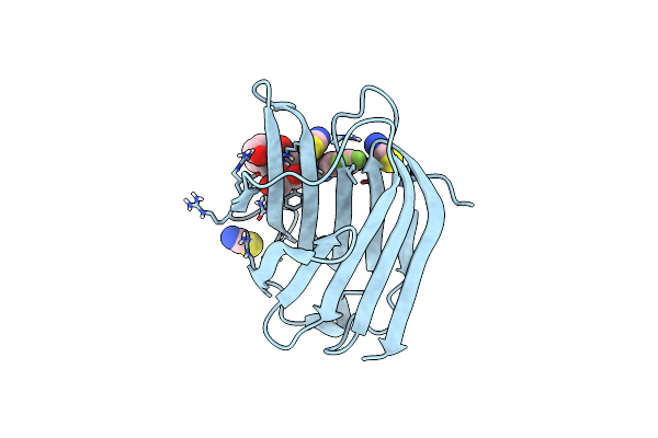 |
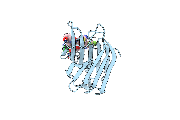
Organism: Homo sapiens
Method: X-RAY DIFFRACTION Release Date: 2025-03-26 Classification: SUGAR BINDING PROTEIN Ligands: SCN, A1IB3 |
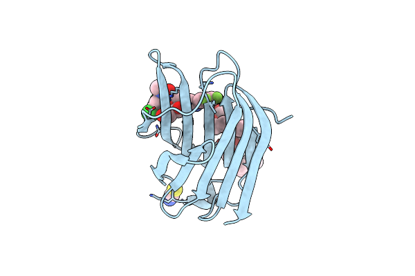 |
Organism: Homo sapiens
Method: X-RAY DIFFRACTION Release Date: 2025-03-26 Classification: SUGAR BINDING PROTEIN Ligands: 2PE, A1IB4, SCN |
 |
Synchrotron X-Ray Crystal Structure Of Oxygen-Bound F87A/F393H P450Bm3 With Decoy C7Prophe (N-Enanthyl-L-Prolyl-L-Phenylalanine) And Substrate Styrene At 2 Mgy X-Ray Dose
Organism: Priestia megaterium nbrc 15308 = atcc 14581
Method: X-RAY DIFFRACTION Resolution:1.60 Å Release Date: 2025-03-26 Classification: OXIDOREDUCTASE Ligands: HEM, SYN, OXY, D0L, GOL |
 |
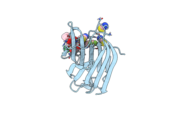
Organism: Homo sapiens
Method: X-RAY DIFFRACTION Resolution:1.10 Å Release Date: 2025-03-12 Classification: SUGAR BINDING PROTEIN Ligands: A1H5A, SCN |
 |
Crystal Structure Of Acyl-Coa Dehydrogenase Fade2 Mutant (Pa0508 M130G E296A Q303A) From Pseudomonas Aeruginosa
Organism: Pseudomonas aeruginosa
Method: X-RAY DIFFRACTION Resolution:1.62 Å Release Date: 2025-02-19 Classification: OXIDOREDUCTASE Ligands: FAD, GOL, PGE |
 |
Crystal Structure Of The Acyl-Coa Dehydrogenase Fade1(Pa0506) E441A From Pseudomonas Aeruginosa Complexed With C16Coa
Organism: Pseudomonas aeruginosa
Method: X-RAY DIFFRACTION Resolution:1.44 Å Release Date: 2025-02-12 Classification: OXIDOREDUCTASE Ligands: PKZ, EDO, PGE, NO3, TRS |
 |
Xfel Crystal Structure Of The Oxidized Form Of F393H P450Bm3 With N-Enanthyl-L-Prolyl-L-Phenylalanine In Complex With Styrene
Organism: Priestia megaterium nbrc 15308 = atcc 14581
Method: X-RAY DIFFRACTION Resolution:1.80 Å Release Date: 2025-02-12 Classification: OXIDOREDUCTASE Ligands: HEM, D0L, SYN, GOL |
 |
Xfel Crystal Structure Of The Oxidized Form Of F87A/F393H P450Bm3 With N-Enanthyl-L-Prolyl-L-Phenylalanine In Complex With Styrene
Organism: Priestia megaterium nbrc 15308 = atcc 14581
Method: X-RAY DIFFRACTION Resolution:1.85 Å Release Date: 2025-02-12 Classification: OXIDOREDUCTASE Ligands: HEM, D0L, SYN, GOL |
 |
Xfel Crystal Structure Of The Reduced Form Of F393H P450Bm3 With N-Enanthyl-L-Prolyl-L-Phenylalanine In Complex With Styrene
Organism: Priestia megaterium nbrc 15308 = atcc 14581
Method: X-RAY DIFFRACTION Resolution:1.60 Å Release Date: 2025-02-12 Classification: OXIDOREDUCTASE Ligands: HEM, D0L, SYN, GOL |
 |
Xfel Crystal Structure Of The Reduced Form Of F87A/F393H P450Bm3 With N-Enanthyl-L-Prolyl-L-Phenylalanine In Complex With Styrene
Organism: Priestia megaterium nbrc 15308 = atcc 14581
Method: X-RAY DIFFRACTION Resolution:1.60 Å Release Date: 2025-02-12 Classification: OXIDOREDUCTASE Ligands: HEM, D0L, SYN, GOL |
 |
Xfel Crystal Structure Of The Oxygen-Bound Form Of F393H P450Bm3 With N-Enanthyl-L-Prolyl-L-Phenylalanine In Complex With Styrene
Organism: Priestia megaterium nbrc 15308 = atcc 14581
Method: X-RAY DIFFRACTION Resolution:1.50 Å Release Date: 2025-02-12 Classification: OXIDOREDUCTASE Ligands: HEM, D0L, SYN, OXY, GOL |
 |
Xfel Crystal Structure Of The Oxygen-Bound Form Of F87A/F393H P450Bm3 With N-Enanthyl-L-Prolyl-L-Phenylalanine In Complex With Styrene
Organism: Priestia megaterium nbrc 15308 = atcc 14581
Method: X-RAY DIFFRACTION Resolution:1.50 Å Release Date: 2025-02-12 Classification: OXIDOREDUCTASE Ligands: HEM, D0L, SYN, OXY, GOL |
 |
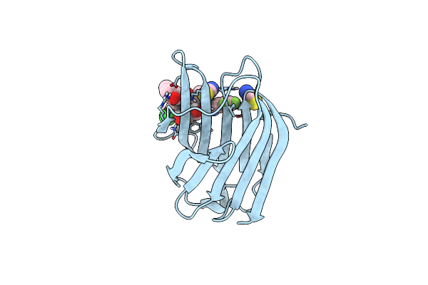
Organism: Homo sapiens
Method: X-RAY DIFFRACTION Release Date: 2025-01-29 Classification: SUGAR BINDING PROTEIN Ligands: A1H1W, SCN |
 |
Organism: Homo sapiens
Method: X-RAY DIFFRACTION Release Date: 2025-01-29 Classification: SUGAR BINDING PROTEIN Ligands: A1H1Y, SCN |
 |
Organism: Homo sapiens
Method: X-RAY DIFFRACTION Release Date: 2025-01-29 Classification: SUGAR BINDING PROTEIN Ligands: A1H1X, SCN |
 |
Organism: Homo sapiens
Method: X-RAY DIFFRACTION Resolution:1.60 Å Release Date: 2025-01-29 Classification: SUGAR BINDING PROTEIN Ligands: A1H2T, SCN, MLI, CL |
 |
Organism: Homo sapiens
Method: X-RAY DIFFRACTION Resolution:1.50 Å Release Date: 2025-01-29 Classification: SUGAR BINDING PROTEIN Ligands: A1H2S |