
Search Count: 6
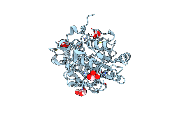 |
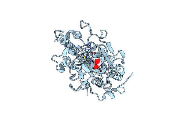
Organism: Escherichia coli
Method: X-RAY DIFFRACTION Resolution:1.20 Å Release Date: 2020-06-03 Classification: ANTIMICROBIAL PROTEIN Ligands: ZN, GOL, BR |
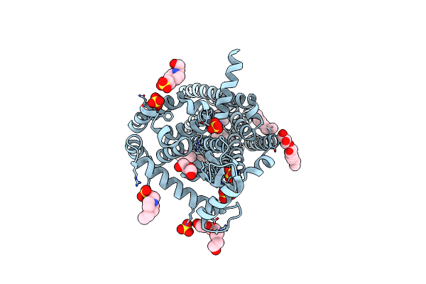 |
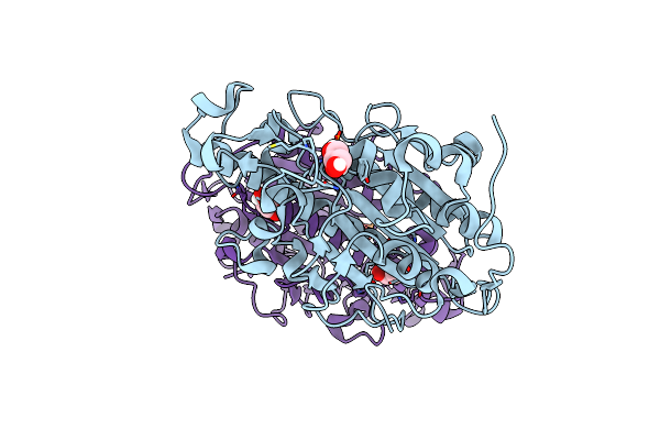
Organism: Homo sapiens, Enterobacteria phage t4
Method: X-RAY DIFFRACTION Resolution:2.80 Å Release Date: 2019-01-30 Classification: MEMBRANE PROTEIN Ligands: 9JU, OLA, OLC, PEG, EPE, PG4, SO4 |
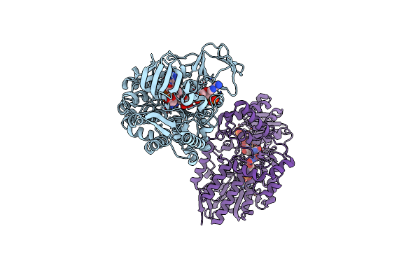 |
Crystal Structure Of Cyclohexanone Monooxygenase From Rhodococcus Sp. Phi1 Bound To Nadp+
Organism: Rhodococcus sp. phi1
Method: X-RAY DIFFRACTION Resolution:2.37 Å Release Date: 2018-09-26 Classification: FLAVOPROTEIN Ligands: FAD, NAP |
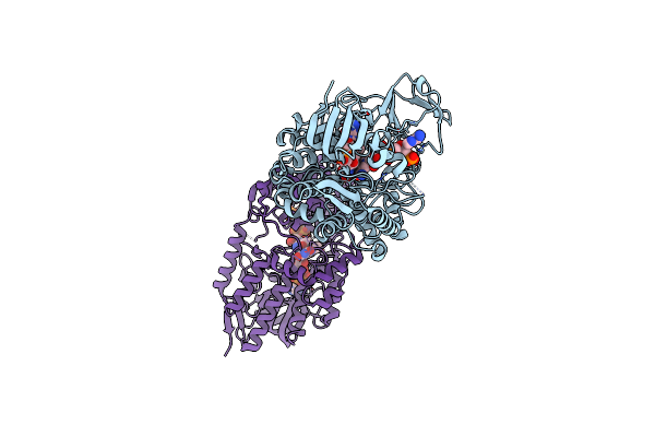 |
Crystal Structure Of Cyclohexanone Monooxygenase Mutant (F249A, F280A And F435A) From Rhodococcus Sp. Phi1 Bound To Nadp+
Organism: Rhodococcus sp. phi1
Method: X-RAY DIFFRACTION Release Date: 2018-09-26 Classification: FLAVOPROTEIN Ligands: FAD, NAP |
 |
Organism: Escherichia coli
Method: X-RAY DIFFRACTION Resolution:1.75 Å Release Date: 2016-12-07 Classification: TRANSFERASE Ligands: ZN, GOL |
 |
Organism: Escherichia coli
Method: X-RAY DIFFRACTION Resolution:1.55 Å Release Date: 2016-12-07 Classification: TRANSFERASE Ligands: ZN, GOL |