
Search Count: 24
 |
Organism: Homo sapiens
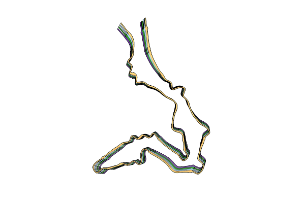
Method: ELECTRON MICROSCOPY Release Date: 2024-09-11 Classification: PROTEIN FIBRIL |
 |
Organism: Homo sapiens
Method: ELECTRON MICROSCOPY Release Date: 2024-09-11 Classification: PROTEIN FIBRIL |
 |
Organism: Homo sapiens
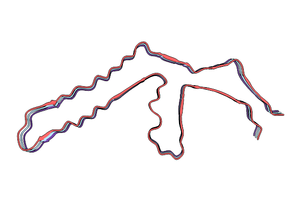
Method: ELECTRON MICROSCOPY Release Date: 2024-09-11 Classification: PROTEIN FIBRIL |
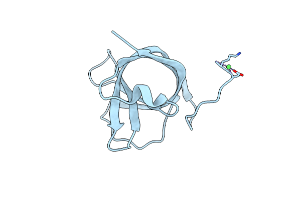 |
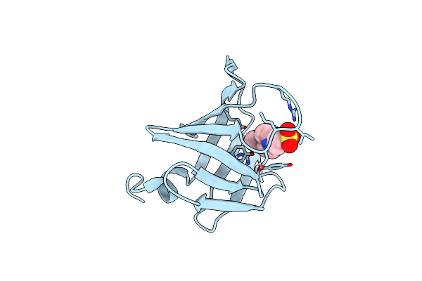
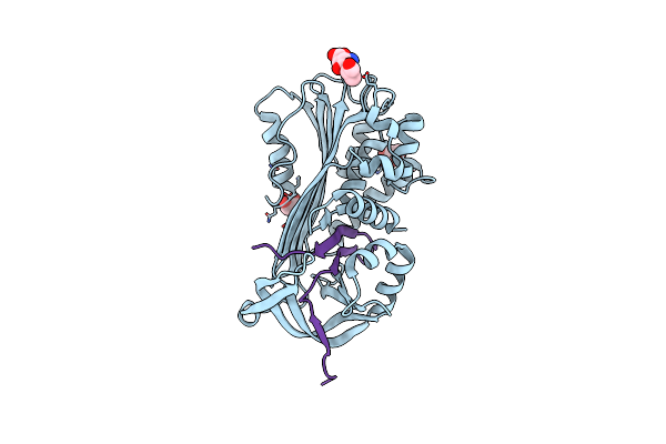
Matrix-Metallopeptidase Inhibitor Potempin A (Pota) From Tannerella Forsythia
Organism: Tannerella forsythia
Method: X-RAY DIFFRACTION Resolution:1.70 Å Release Date: 2022-12-21 Classification: HYDROLASE INHIBITOR Ligands: NI |
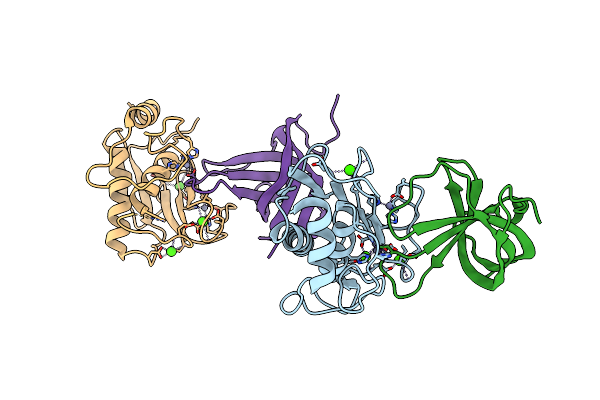 |
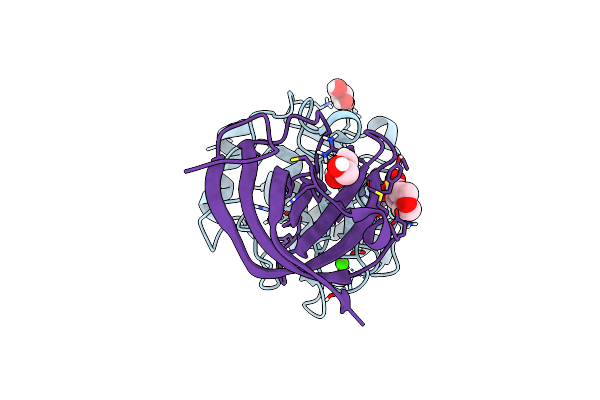
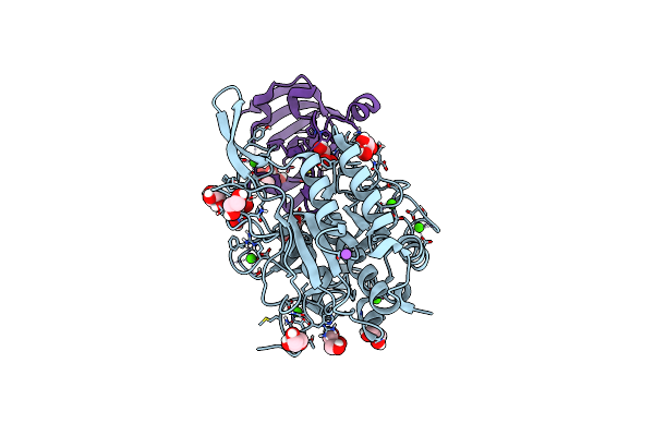
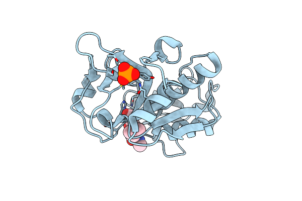
Potempin A (Pota) From Tannerella Forsythia In Complex With The Catalytic Domain Of Human Mmp-12
Organism: Homo sapiens, Tannerella forsythia
Method: X-RAY DIFFRACTION Resolution:1.85 Å Release Date: 2022-12-21 Classification: HYDROLASE INHIBITOR Ligands: CA, ZN |
 |
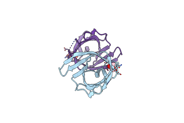
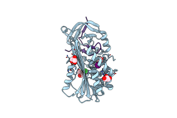
Matrix-Metallopeptidase Inhibitor Potempin A (Pota) From Tannerella Forsythia In Complex With T. Forsythia Karilysin.
Organism: Tannerella forsythia
Method: X-RAY DIFFRACTION Resolution:1.35 Å Release Date: 2022-12-21 Classification: HYDROLASE INHIBITOR Ligands: ZN, CA, MES, GOL |
 |
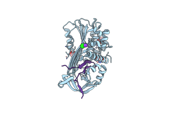
Structure Of Tannerella Forsythia Selenomethionine-Derivatized Potempin D Mutant I53M
Organism: Tannerella forsythia (strain atcc 43037 / jcm 10827 / ccug 21028 a / kctc 5666 / fdc 338)
Method: X-RAY DIFFRACTION Resolution:2.40 Å Release Date: 2022-12-21 Classification: HYDROLASE INHIBITOR Ligands: EDO |
 |
Organism: Tannerella forsythia
Method: X-RAY DIFFRACTION Resolution:2.00 Å Release Date: 2022-12-21 Classification: HYDROLASE INHIBITOR Ligands: GOL |
 |
Organism: Tannerella forsythia
Method: X-RAY DIFFRACTION Resolution:1.80 Å Release Date: 2022-12-21 Classification: HYDROLASE INHIBITOR Ligands: EPE |
 |
Organism: Tannerella forsythia
Method: X-RAY DIFFRACTION Resolution:1.10 Å Release Date: 2022-12-21 Classification: HYDROLASE INHIBITOR |
 |
Organism: Tannerella forsythia
Method: X-RAY DIFFRACTION Resolution:3.00 Å Release Date: 2017-05-24 Classification: HYDROLASE INHIBITOR |
 |
Structure Of The Trypsin Induced Serpin-Type Proteinase Inhibitor, Miropin.
Organism: Tannerella forsythia
Method: X-RAY DIFFRACTION Resolution:1.60 Å Release Date: 2017-05-24 Classification: Hydrolase inhibitor Ligands: ASP, SER, GOL |
 |
Structure Of The Subtilisin Induced Serpin-Type Proteinase Inhibitor, Miropin.
Organism: Tannerella forsythia
Method: X-RAY DIFFRACTION Resolution:1.70 Å Release Date: 2017-05-24 Classification: HYDROLASE INHIBITOR Ligands: K, IOD, CL, GOL |
 |
Structure Of The Trypsin Induced Serpin-Type Proteinase Inhibitor, Miropin (V367K/K368A Mutant).
Organism: Tannerella forsythia
Method: X-RAY DIFFRACTION Resolution:1.50 Å Release Date: 2017-05-24 Classification: HYDROLASE Ligands: ZN, K, IOD, CL, TRS, GOL |
 |
Crystal Structure Of Dionain-1, The Major Endopeptidase In The Venus Flytrap Digestive Juice
Organism: Dionaea muscipula
Method: X-RAY DIFFRACTION Resolution:1.50 Å Release Date: 2015-12-09 Classification: HYDROLASE Ligands: E64, PO4 |
 |
Structure Of Ragb, A Major Immunodominant Virulence Factor Of Porphyromonas Gingivalis.
Organism: Porphyromonas gingivalis (strain atcc baa-308 / w83)
Method: X-RAY DIFFRACTION Resolution:2.40 Å Release Date: 2015-10-21 Classification: MEMBRANE PROTEIN Ligands: 3DO, TG6, GOL, ACT, RP3 |
 |
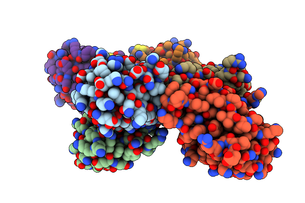
Cryo-Em Single Particle 3D Reconstruction Of The Native Conformation Of E. Coli Alpha-2-Macroglobulin (Ecam)
Organism: Escherichia coli k-12
Method: ELECTRON MICROSCOPY Resolution:16.00 Å Release Date: 2015-07-29 Classification: HYDROLASE INHIBITOR |
 |
Crystal Structure Of Porphyromonas Gingivalis Peptidylarginine Deiminase (Ppad) Substrate-Unbound.
Organism: Porphyromonas gingivalis w83
Method: X-RAY DIFFRACTION Resolution:1.50 Å Release Date: 2015-07-15 Classification: HYDROLASE |
 |
Crystal Structure Of Porphyromonas Gingivalis Peptidylarginine Deiminase (Ppad) In Complex With Dipeptide Asp-Gln.
Organism: Porphyromonas gingivalis
Method: X-RAY DIFFRACTION Resolution:1.40 Å Release Date: 2015-07-01 Classification: HYDROLASE |
 |
Crystal Structure Of Porphyromonas Gingivalis Peptidylarginine Deiminase (Ppad) Mutant C351A In Complex With Dipeptide Met-Arg.
Organism: Porphyromonas gingivalis (strain atcc baa-308 / w83)
Method: X-RAY DIFFRACTION Resolution:1.80 Å Release Date: 2015-07-01 Classification: HYDROLASE |