
Search Count: 39
 |
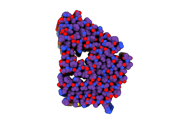
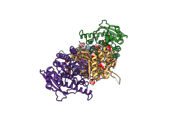
Organism: Homo sapiens, Pyrococcus abyssi
Method: ELECTRON MICROSCOPY Release Date: 2025-09-24 Classification: MEMBRANE PROTEIN Ligands: 5YM, A1EM1, 1DO, A1EM3, A1EQ8, A1EM2 |
 |
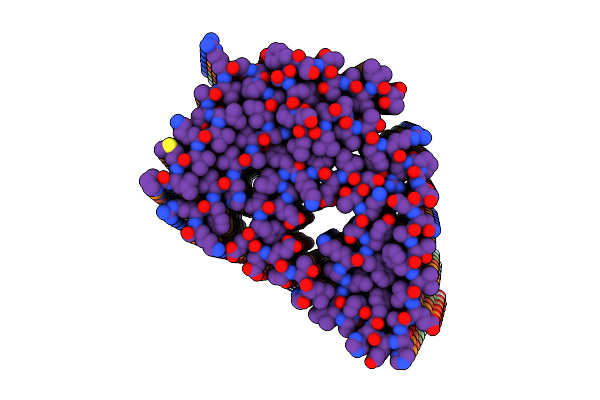
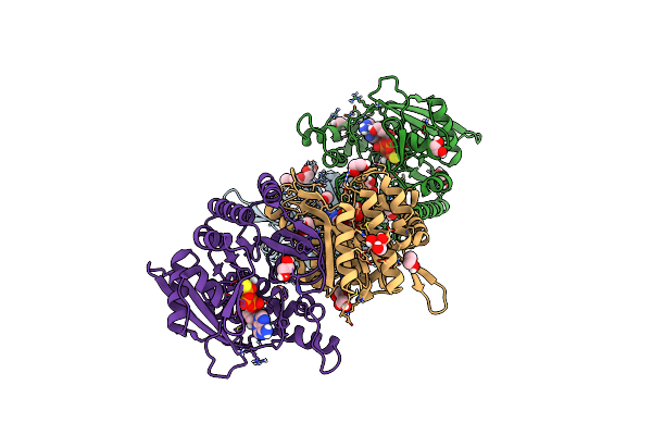
Organism: Escherichia coli, Homo sapiens
Method: ELECTRON MICROSCOPY Release Date: 2025-09-24 Classification: MEMBRANE PROTEIN Ligands: 5YM, 1DO, A1AJD, A1EM2 |
 |
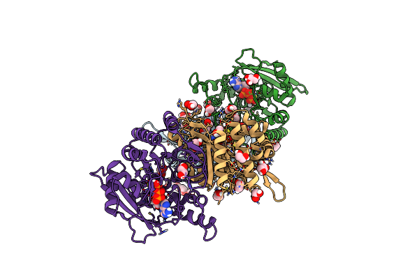
Organism: Escherichia coli, Homo sapiens
Method: ELECTRON MICROSCOPY Release Date: 2025-09-24 Classification: MEMBRANE PROTEIN Ligands: A1EM2, 1DO |
 |
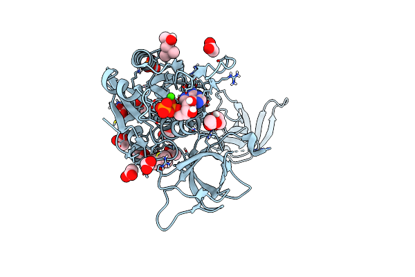
Crystal Structure Of P110Alpha-Ras Binding Domain (Rbd) In Complex With Molecular Glue D927
Organism: Homo sapiens
Method: X-RAY DIFFRACTION Release Date: 2025-07-23 Classification: SIGNALING PROTEIN Ligands: A1ATF, MG, MRD, MPD |
 |
Crystal Structure Of P110Alpha-Ras Binding Domain (Rbd) In Complex With Molecular Glue D223
Organism: Homo sapiens
Method: X-RAY DIFFRACTION Release Date: 2025-07-23 Classification: SIGNALING PROTEIN Ligands: A1AZD |
 |
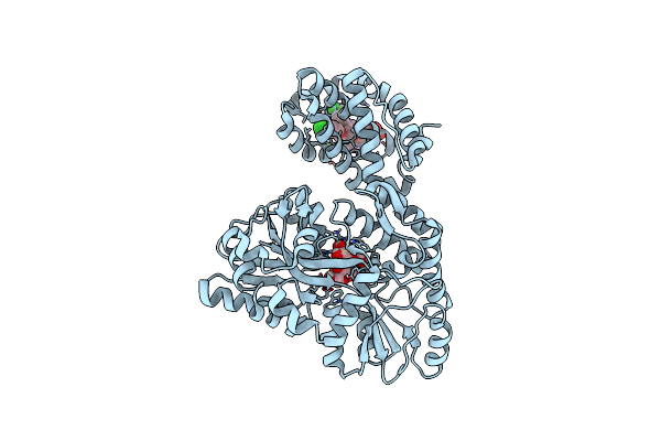
Crystal Structure Of The Kras-P110Alpha Complex In The Presence Of Molecular Glue D223
Organism: Homo sapiens
Method: X-RAY DIFFRACTION Release Date: 2025-07-23 Classification: ONCOPROTEIN Ligands: A1AZD, GOL, MG, GNP |
 |
Organism: Homo sapiens
Method: X-RAY DIFFRACTION Resolution:1.90 Å Release Date: 2024-08-07 Classification: APOPTOSIS Ligands: A1ALT |
 |
Organism: Homo sapiens
Method: ELECTRON MICROSCOPY Release Date: 2024-06-26 Classification: PROTEIN FIBRIL |
 |
Organism: Homo sapiens
Method: ELECTRON MICROSCOPY Release Date: 2024-06-26 Classification: PROTEIN FIBRIL |
 |
Organism: Homo sapiens
Method: ELECTRON MICROSCOPY Release Date: 2023-12-06 Classification: PROTEIN FIBRIL |
 |
Organism: Homo sapiens
Method: ELECTRON MICROSCOPY Release Date: 2023-12-06 Classification: PROTEIN FIBRIL |
 |
Organism: Homo sapiens
Method: ELECTRON MICROSCOPY Release Date: 2023-12-06 Classification: PROTEIN FIBRIL |
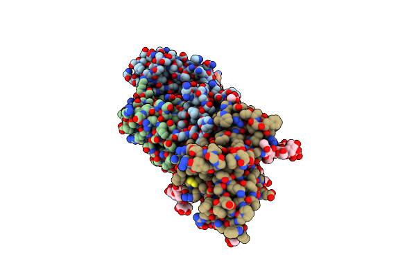 |
Crystal Structure Of Broadly Neutralizing Antibody Mab1198 In Complex With Hepatitis C Virus Envelope Glycoprotein E2 Ectodomain
Organism: Homo sapiens, Hepacivirus c
Method: X-RAY DIFFRACTION Resolution:2.70 Å Release Date: 2022-01-12 Classification: VIRAL PROTEIN/IMMUNE SYSTEM Ligands: NAG |
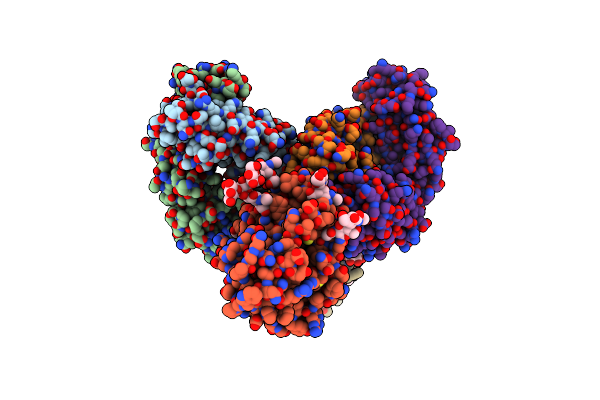 |
Crystal Structure Of Broadly Neutralizing Antibody Mab1382 In Complex With Hepatitis C Virus Envelope Glycoprotein E2 Ectodomain
Organism: Homo sapiens, Hepacivirus c
Method: X-RAY DIFFRACTION Resolution:3.24 Å Release Date: 2022-01-12 Classification: VIRAL PROTEIN/IMMUNE SYSTEM Ligands: NAG |
 |
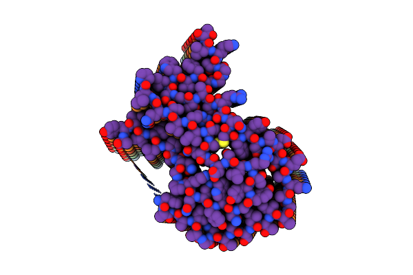
Organism: Mus musculus
Method: X-RAY DIFFRACTION Resolution:2.45 Å Release Date: 2021-04-14 Classification: TRANSFERASE Ligands: PG4, GOL, EDO |
 |
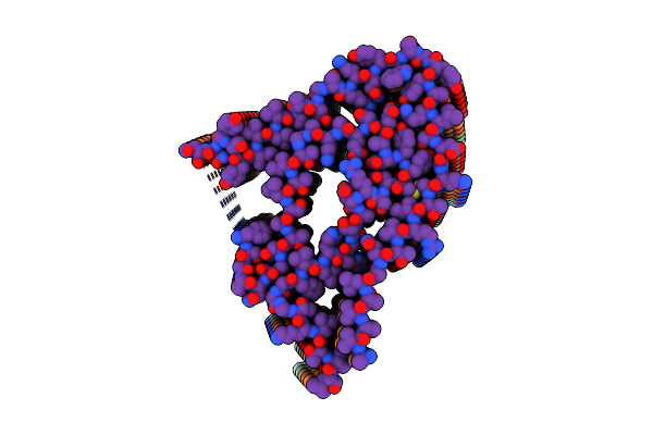
Organism: Mus musculus
Method: X-RAY DIFFRACTION Resolution:2.90 Å Release Date: 2021-04-14 Classification: TRANSFERASE Ligands: PG4, AGS, EDO, GOL |
 |
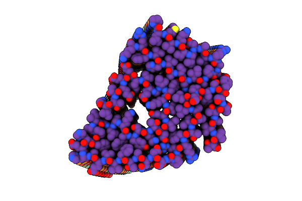
Crystal Structure Of Mouse Pyridoxal Kinase In Complex With Atp-Gamma-S And Artesunate
Organism: Mus musculus
Method: X-RAY DIFFRACTION Resolution:2.40 Å Release Date: 2021-04-14 Classification: TRANSFERASE Ligands: GOL, PG4, D95, AGS, EDO |
 |
Organism: Homo sapiens, Ebola virus
Method: ELECTRON MICROSCOPY Release Date: 2019-10-02 Classification: VIRAL PROTEIN Ligands: MAN, NAG |
 |
Organism: Rattus norvegicus
Method: X-RAY DIFFRACTION Resolution:1.50 Å Release Date: 2019-01-16 Classification: STRUCTURAL PROTEIN Ligands: CA, ADP, PO4, MPD, MRD, ACT, D95, CL |
 |
Organism: Rattus norvegicus
Method: X-RAY DIFFRACTION Resolution:1.50 Å Release Date: 2019-01-16 Classification: STRUCTURAL PROTEIN Ligands: CA, PO4, ACT, NA, ADP, MPD, D8Z |