
Search Count: 359
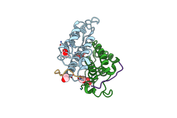 |
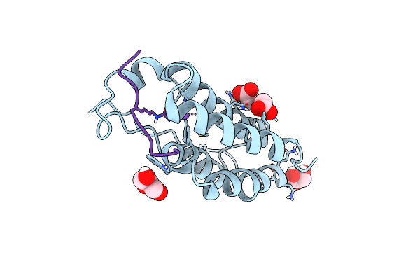
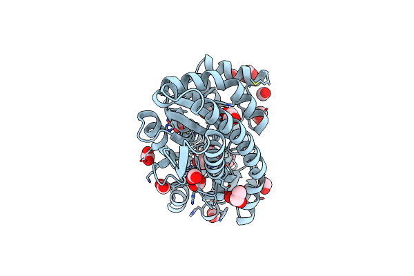
Crystal Structure Of Human Bromodomain Containing Protein 3 (Brd3) In Complex With Hnrnpk
Organism: Homo sapiens
Method: X-RAY DIFFRACTION Resolution:1.85 Å Release Date: 2022-08-03 Classification: SIGNALING PROTEIN Ligands: EDO, SO4, GOL |
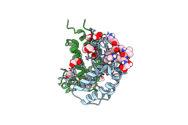 |
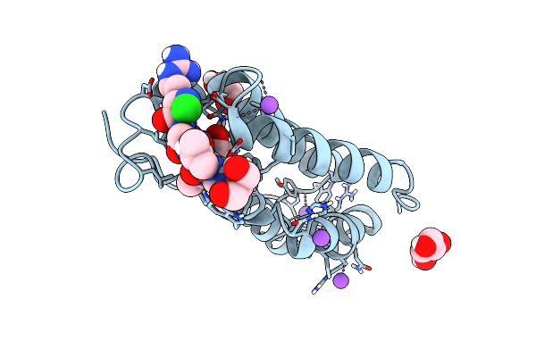
Crystal Structure Of Human Bromodomain Containing Protein 3 (Brd3) In Complex With Shmt
Organism: Homo sapiens
Method: X-RAY DIFFRACTION Resolution:1.50 Å Release Date: 2022-08-03 Classification: SIGNALING PROTEIN Ligands: EDO, GOL |
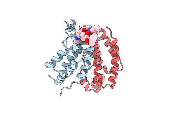 |
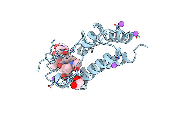
Crystal Structure Of Human Bromodomain Containing Protein 3 (Brd3) In Complex With Ilf3
Organism: Homo sapiens
Method: X-RAY DIFFRACTION Resolution:2.10 Å Release Date: 2022-08-03 Classification: SIGNALING PROTEIN Ligands: EDO |
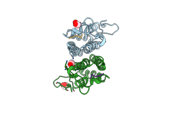 |
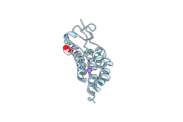
Crystal Structure Of Human Bromodomain Containing Protein 3 (Brd3) In Complex With Bcltf1
Organism: Homo sapiens
Method: X-RAY DIFFRACTION Resolution:1.95 Å Release Date: 2022-08-03 Classification: SIGNALING PROTEIN Ligands: EDO |
 |
Crystal Structure Of Human Bromodomain Containing Protein 4 (Brd4) In Complex With Hnrnpk
Organism: Homo sapiens
Method: X-RAY DIFFRACTION Resolution:1.38 Å Release Date: 2022-08-03 Classification: SIGNALING PROTEIN |
 |
Crystal Structure Of Human Bromodomain Containing Protein 4 (Brd4) In Complex With Shmt
Organism: Homo sapiens
Method: X-RAY DIFFRACTION Resolution:1.25 Å Release Date: 2022-08-03 Classification: SIGNALING PROTEIN Ligands: EDO, NA, CL, GOL |
 |
Crystal Structure Of Human Bromodomain Containing Protein 4 (Brd4) In Complex With Ilf3
Organism: Homo sapiens
Method: X-RAY DIFFRACTION Resolution:1.72 Å Release Date: 2022-08-03 Classification: SIGNALING PROTEIN |
 |
Crystal Structure Of Human Bromodomain Containing Protein 4 (Brd4) In Complex With Bcltf1
Organism: Homo sapiens
Method: X-RAY DIFFRACTION Resolution:1.45 Å Release Date: 2022-08-03 Classification: SIGNALING PROTEIN Ligands: EDO, NA |
 |
Organism: Homo sapiens
Method: X-RAY DIFFRACTION Resolution:3.50 Å Release Date: 2021-10-27 Classification: IMMUNE SYSTEM |
 |
Organism: Mus musculus
Method: X-RAY DIFFRACTION Resolution:2.10 Å Release Date: 2021-10-27 Classification: IMMUNE SYSTEM Ligands: SO4 |
 |
Crystal Structure Of A Marine Metagenome Trap Solute Binding Protein Specific For Pyroglutamate (Sorcerer Ii Global Ocean Sampling Expedition, Unidentified Microbe, Scf7180008839099) In Complex With Co-Purified Pyroglutamate
Organism: Uncultured bacterium
Method: X-RAY DIFFRACTION Resolution:1.40 Å Release Date: 2020-04-29 Classification: SUGAR BINDING PROTEIN Ligands: PCA, EDO, CL, FMT |
 |
Organism: Homo sapiens
Method: X-RAY DIFFRACTION Resolution:2.88 Å Release Date: 2019-07-03 Classification: IMMUNE SYSTEM Ligands: MG |
 |
Organism: Homo sapiens
Method: X-RAY DIFFRACTION Resolution:1.95 Å Release Date: 2019-07-03 Classification: IMMUNE SYSTEM |
 |
Organism: Homo sapiens
Method: X-RAY DIFFRACTION Resolution:1.95 Å Release Date: 2019-07-03 Classification: IMMUNE SYSTEM |
 |
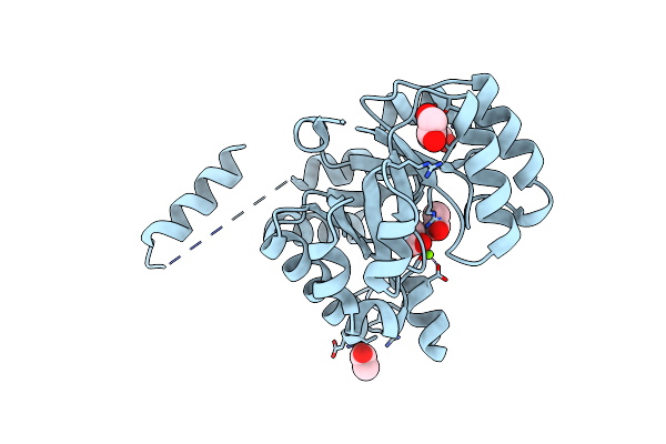
Crystal Structure Of Protein Cite From Mycobacterium Tuberculosis In Complex With Magnesium, Pyruvate And Citramalyl-Coa
Organism: Mycobacterium tuberculosis h37rv
Method: X-RAY DIFFRACTION Resolution:1.83 Å Release Date: 2018-08-01 Classification: LYASE Ligands: MG, CQM, PO4, GOL, CL, PYR |
 |
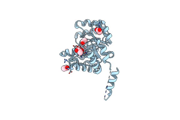
Crystal Structure Of Protein Cite From Mycobacterium Tuberculosis In Complex With Magnesium, Pyruvate And Coenzyme A
Organism: Mycobacterium tuberculosis h37rv
Method: X-RAY DIFFRACTION Resolution:1.72 Å Release Date: 2018-08-01 Classification: LYASE Ligands: MG, PYR, COA, PO4, GOL |
 |
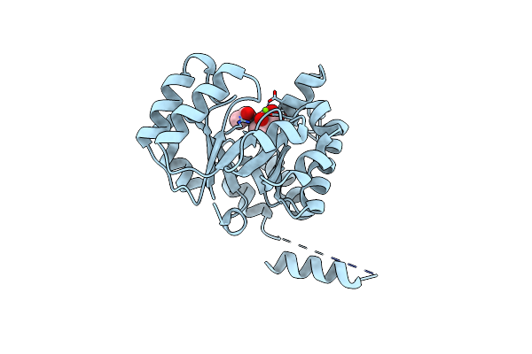
Crystal Structure Of Protein Cite From Mycobacterium Tuberculosis In Complex With Magnesium, Acetoacetate And Coenzyme A
Organism: Mycobacterium tuberculosis h37rv
Method: X-RAY DIFFRACTION Resolution:2.04 Å Release Date: 2018-08-01 Classification: LYASE Ligands: MG, COA, AAE, SO4, GOL |
 |
Crystal Structure Of Protein Cite From Mycobacterium Tuberculosis In Complex With Magnesium And Acetate
Organism: Mycobacterium tuberculosis h37rv
Method: X-RAY DIFFRACTION Resolution:1.61 Å Release Date: 2018-08-01 Classification: LYASE Ligands: MG, ACT, EDO |
 |
Crystal Structure Of Protein Cite From Mycobacterium Tuberculosis In Complex With Magnesium And Pyruvate
Organism: Mycobacterium tuberculosis h37rv
Method: X-RAY DIFFRACTION Resolution:1.73 Å Release Date: 2018-08-01 Classification: LYASE Ligands: MG, ACT, PYR |
 |
Crystal Structure Of Protein Cite From Mycobacterium Tuberculosis In Complex With Magnesium And Acetoacetate
Organism: Mycobacterium tuberculosis h37rv
Method: X-RAY DIFFRACTION Resolution:1.73 Å Release Date: 2018-08-01 Classification: LYASE Ligands: MG, ACT, AAE |