
Search Count: 28
 |
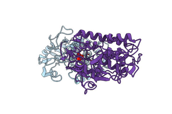
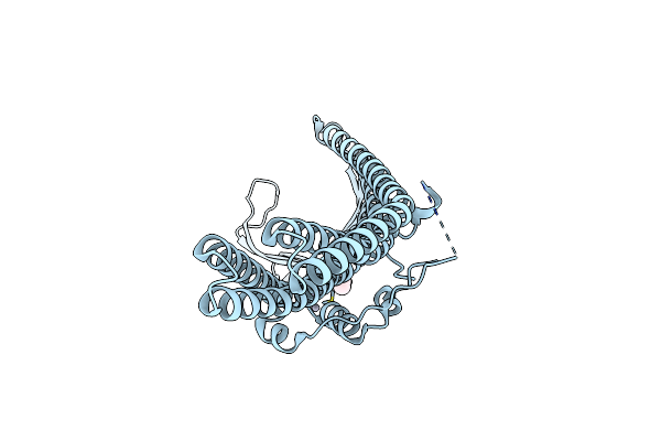
Organism: Pseudomonas aeruginosa pao1
Method: X-RAY DIFFRACTION Resolution:2.90 Å Release Date: 2019-06-26 Classification: RNA BINDING PROTEIN Ligands: ZN, GDP |
 |
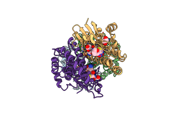
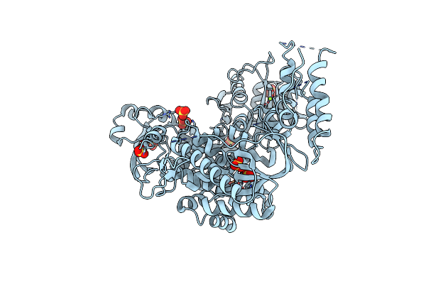
Organism: natrialba magadii atcc 43099
Method: X-RAY DIFFRACTION Resolution:2.61 Å Release Date: 2019-02-13 Classification: TRANSFERASE Ligands: SO4 |
 |
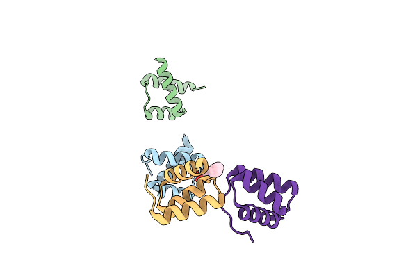
Organism: Homo sapiens
Method: SOLUTION NMR Release Date: 2012-06-27 Classification: DNA BINDING PROTEIN, CHAPERONE |
 |
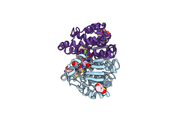
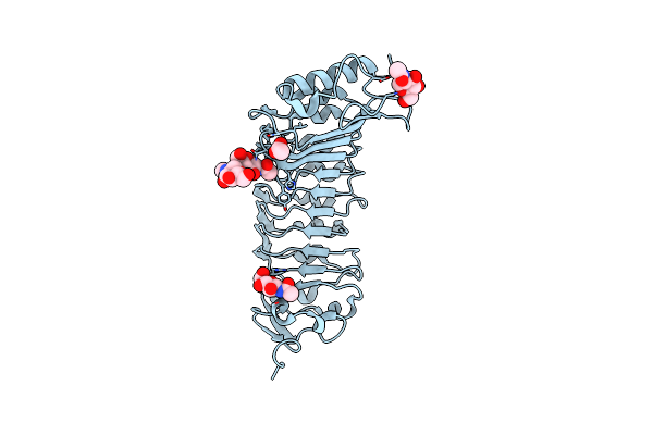
Crystal Structure Of Mu Class Glutathione S-Transferase (Gstm2-2) In Complex With Glutathione And 6-(7-Nitro-2,1,3-Benzoxadiazol-4-Ylthio)Hexanol (Nbdhex)
Organism: Homo sapiens
Method: X-RAY DIFFRACTION Resolution:2.50 Å Release Date: 2009-10-27 Classification: TRANSFERASE/TRANSFERASE INHIBITOR Ligands: BYG, GSH, EDO |
 |
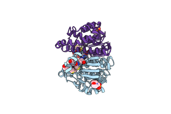
Crystal Strcture Of Human Pi Class Glutathione S-Transferase Gstp1-1 In Complex With 6-(7-Nitro-2,1,3-Benzoxadiazol-4-Ylthio)Hexanol (Nbdhex)
Organism: Homo sapiens
Method: X-RAY DIFFRACTION Resolution:1.53 Å Release Date: 2009-10-27 Classification: TRANSFERASE/TRANSFERASE INHIBITOR Ligands: GSH, N11, MES, SO4 |
 |
Structural Basis For The Binding Of The Anti-Cancer Compound 6-(7-Nitro-2,1,3-Benzoxadiazol-4-Ylthio)Hexanol (Nbdhex) To Human Glutathione S-Transferases
Organism: Homo sapiens
Method: X-RAY DIFFRACTION Resolution:1.80 Å Release Date: 2009-10-27 Classification: TRANSFERASE/TRANSFERASE INHIBITOR Ligands: GSH, N11, MES |
 |
Crystal Structure Of Ochrobactrum Anthropi Glutathione Transferase Cys10Ala Mutant With Glutathione Bound At The H-Site
Organism: Ochrobactrum anthropi
Method: X-RAY DIFFRACTION Resolution:1.80 Å Release Date: 2008-01-15 Classification: TRANSFERASE Ligands: SO4, GSH |
 |
Crystal Structure Of The Polygalacturonase From Colletotrichum Lupini And Its Implications For The Interaction With Polygalacturonase-Inhibiting Proteins
Organism: Colletotrichum lupini
Method: X-RAY DIFFRACTION Resolution:1.94 Å Release Date: 2007-10-23 Classification: HYDROLASE Ligands: PG4, PEG, ACY |
 |
Structure Of The Glutathione Transferase From Ochrobactrum Anthropi In Complex With Glutathione
Organism: Ochrobactrum anthropi
Method: X-RAY DIFFRACTION Resolution:2.10 Å Release Date: 2007-09-25 Classification: TRANSFERASE Ligands: SO4, GSH |
 |
The Crystal Structure Of The Bar Domain From Human Bin1/Amphiphysin Ii And Its Implications For Molecular Recognition
Organism: Homo sapiens
Method: X-RAY DIFFRACTION Resolution:1.99 Å Release Date: 2006-11-14 Classification: ENDOCYTOSIS/EXOCYTOSIS, MEMBRANE PROTEIN Ligands: XE |
 |
The Crystal Structure Of The Outer Membrane Protein Vcec From The Bacterial Pathogen Vibrio Cholerae At 1.8 Resolution
Organism: Vibrio cholerae
Method: X-RAY DIFFRACTION Resolution:1.80 Å Release Date: 2005-03-01 Classification: MEMBRANE PROTEIN Ligands: BOG, HG |
 |
Insight In Dna Replication: The Crystal Structure Of Dna Polymerase B1 From The Archaeon Sulfolobus Solfataricus
Organism: Sulfolobus solfataricus
Method: X-RAY DIFFRACTION Resolution:2.40 Å Release Date: 2004-11-09 Classification: TRANSFERASE Ligands: SO4, MG |
 |
Organism: Drosophila melanogaster
Method: X-RAY DIFFRACTION Resolution:2.10 Å Release Date: 2003-10-14 Classification: DNA BINDING PROTEIN Ligands: NHE |
 |
Organism: Drosophila melanogaster
Method: X-RAY DIFFRACTION Resolution:2.10 Å Release Date: 2003-10-14 Classification: DNA BINDING PROTEIN Ligands: NHE |
 |
The Crystal Structure Of Pgip (Polygalacturonase Inhibiting Protein), A Leucine Rich Repeat Protein Involved In Plant Defense
Organism: Phaseolus vulgaris
Method: X-RAY DIFFRACTION Resolution:1.70 Å Release Date: 2003-07-24 Classification: INHIBITOR Ligands: NAG, ACT |
 |
Organism: Physeter catodon
Method: X-RAY DIFFRACTION Resolution:1.80 Å Release Date: 2003-06-10 Classification: OXYGEN STORAGE/TRANSPORT Ligands: OH, SO4, HEM |
 |
Organism: Physeter catodon
Method: X-RAY DIFFRACTION Resolution:1.80 Å Release Date: 2003-06-10 Classification: OXYGEN STORAGE/TRANSPORT Ligands: OH, SO4, HEM |
 |
Organism: Physeter catodon
Method: X-RAY DIFFRACTION Resolution:1.60 Å Release Date: 2003-06-10 Classification: OXYGEN STORAGE/TRANSPORT Ligands: SO4, OH, HEM |
 |
Organism: Physeter catodon
Method: X-RAY DIFFRACTION Resolution:1.60 Å Release Date: 2003-06-10 Classification: OXYGEN STORAGE/TRANSPORT Ligands: OH, SO4, HEM |
 |
Organism: Physeter catodon
Method: X-RAY DIFFRACTION Resolution:1.04 Å Release Date: 2003-06-10 Classification: oxygen storage/transport Ligands: OH, SO4, HEM |