
Search Count: 10
 |
Organism: Homo sapiens, Severe acute respiratory syndrome coronavirus 2
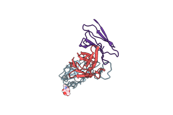
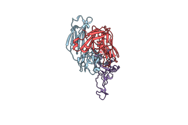
Method: ELECTRON MICROSCOPY Release Date: 2023-11-01 Classification: VIRAL PROTEIN Ligands: NAG |
 |
Organism: Severe acute respiratory syndrome coronavirus 2
Method: ELECTRON MICROSCOPY Release Date: 2023-11-01 Classification: VIRAL PROTEIN Ligands: NAG |
 |
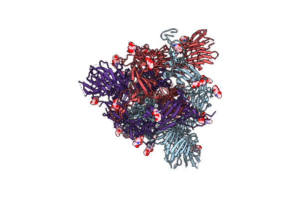
Organism: Homo sapiens, Severe acute respiratory syndrome coronavirus 2
Method: ELECTRON MICROSCOPY Release Date: 2022-08-03 Classification: VIRAL PROTEIN Ligands: NAG |
 |
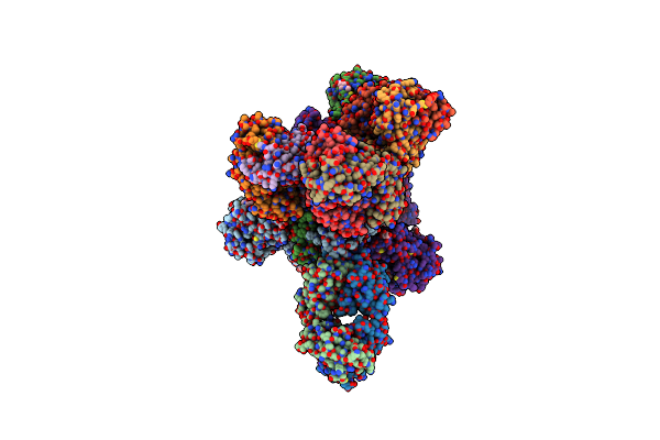
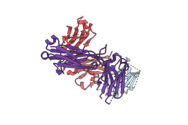
Sars-Cov-2 S Omicron Spike B.1.1.529 - Rbd Up - 1-P2G3 And 1-P5C3 Fabs (Local)
Organism: Homo sapiens, Severe acute respiratory syndrome coronavirus 2
Method: ELECTRON MICROSCOPY Release Date: 2022-08-03 Classification: VIRAL PROTEIN |
 |
Organism: Severe acute respiratory syndrome coronavirus 2, Homo sapiens
Method: ELECTRON MICROSCOPY Release Date: 2022-08-03 Classification: VIRAL PROTEIN |
 |
Organism: Severe acute respiratory syndrome coronavirus 2, Homo sapiens
Method: ELECTRON MICROSCOPY Release Date: 2021-10-13 Classification: VIRAL PROTEIN |
 |
Organism: Severe acute respiratory syndrome coronavirus 2, Homo sapiens
Method: ELECTRON MICROSCOPY Release Date: 2021-10-13 Classification: VIRAL PROTEIN |
 |
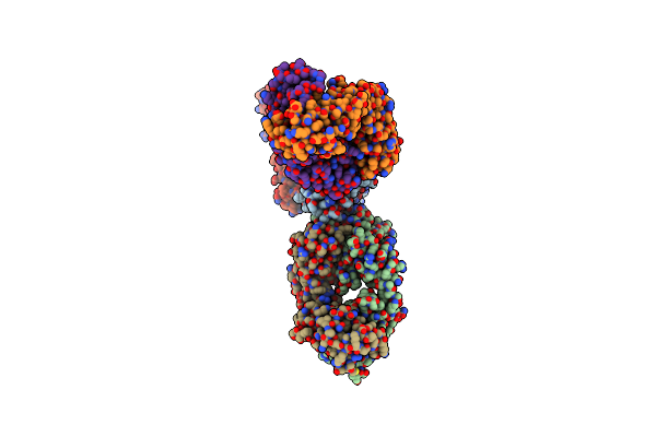
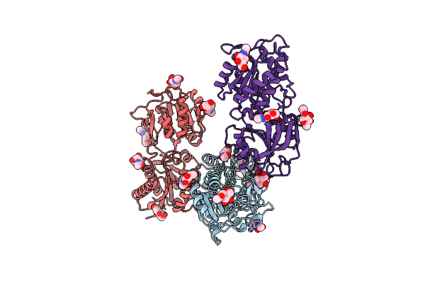
Organism: Mus musculus, Homo sapiens
Method: X-RAY DIFFRACTION Resolution:2.20 Å Release Date: 2019-06-05 Classification: IMMUNE SYSTEM |
 |
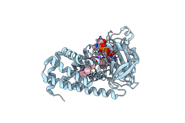
Organism: Rattus norvegicus
Method: X-RAY DIFFRACTION Resolution:3.40 Å Release Date: 2011-03-23 Classification: TRANSPORT PROTEIN Ligands: NAG, CL |
 |
The X-Ray Structure Of Ferric Escherichia Coli Flavohemoglobin Reveals An Unespected Geometry Of The Distal Heme Pocket
Organism: Escherichia coli
Method: X-RAY DIFFRACTION Resolution:2.19 Å Release Date: 2002-08-06 Classification: OXIDOREDUCTASE Ligands: FAD, HEM, NA, CL |