
Search Count: 84
 |
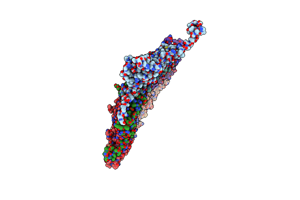
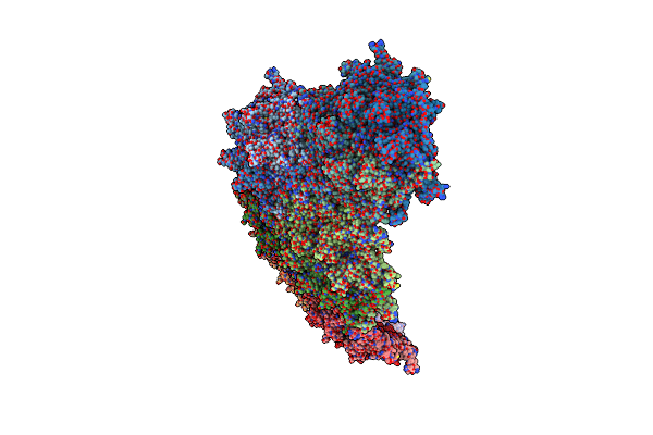
Organism: Bacillus phage spo1
Method: ELECTRON MICROSCOPY Release Date: 2025-08-27 Classification: VIRUS |
 |
Organism: Xanthomonas phage phixacjx1
Method: ELECTRON MICROSCOPY Release Date: 2025-05-07 Classification: VIRUS |
 |
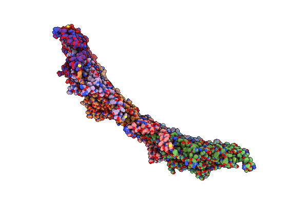
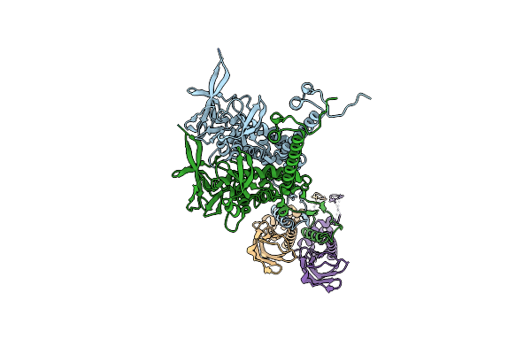
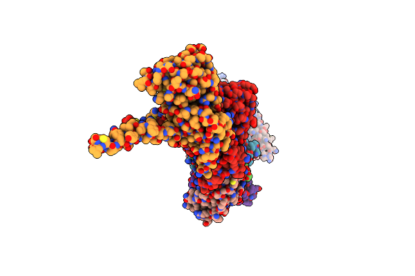
The Composite Cryo-Em Structure Of The Head-To-Tail Connector And Head-Proximal Tail Components Of Bacteriophage Phixacjx1
Organism: Xanthomonas phage phixacjx1
Method: ELECTRON MICROSCOPY Release Date: 2025-05-07 Classification: VIRUS |
 |
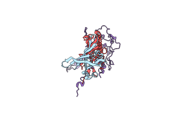
Organism: Mycolicibacterium phage mycofy1
Method: ELECTRON MICROSCOPY Release Date: 2025-04-16 Classification: VIRAL PROTEIN |
 |
Organism: Mycolicibacterium phage mycofy1
Method: ELECTRON MICROSCOPY Release Date: 2025-04-16 Classification: VIRAL PROTEIN |
 |
Organism: Mycolicibacterium phage mycofy1
Method: ELECTRON MICROSCOPY Release Date: 2025-04-16 Classification: VIRAL PROTEIN |
 |
Organism: Mycolicibacterium phage mycofy1
Method: ELECTRON MICROSCOPY Release Date: 2025-04-16 Classification: VIRAL PROTEIN |
 |
Organism: Mycolicibacterium phage mycofy1
Method: ELECTRON MICROSCOPY Release Date: 2025-04-16 Classification: VIRAL PROTEIN |
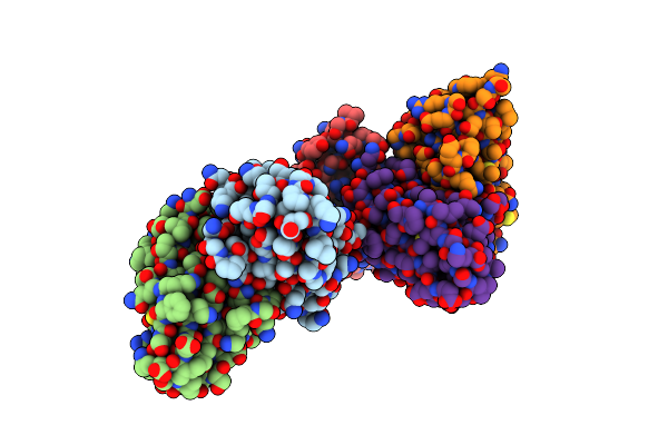 |
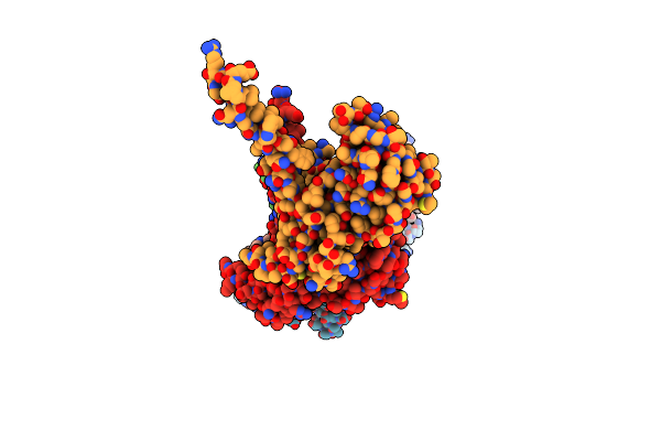
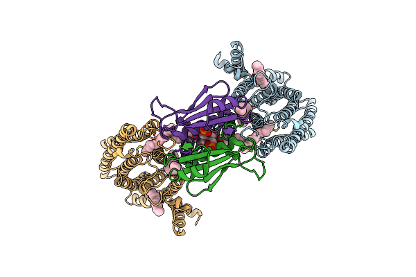
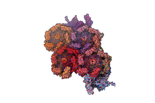
Locally Refined Region Of Sars-Cov-2 Spike In Complex With Antibodies 9G11 And 3E2.
Organism: Severe acute respiratory syndrome coronavirus 2, Mus musculus
Method: ELECTRON MICROSCOPY Release Date: 2025-02-12 Classification: VIRAL PROTEIN/IMMUNE SYSTEM Ligands: NAG |
 |
Organism: Escherichia phage fcwl1
Method: ELECTRON MICROSCOPY Release Date: 2025-01-22 Classification: VIRAL PROTEIN |
 |
Organism: Escherichia phage fcwl1
Method: ELECTRON MICROSCOPY Release Date: 2025-01-22 Classification: VIRUS |
 |
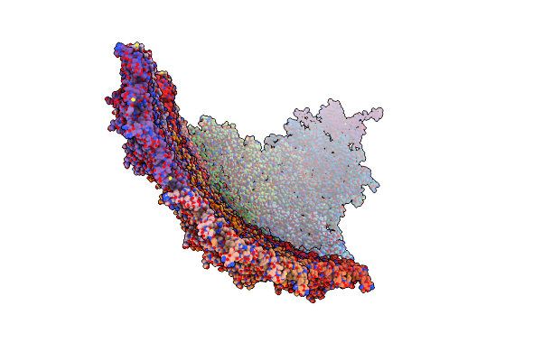
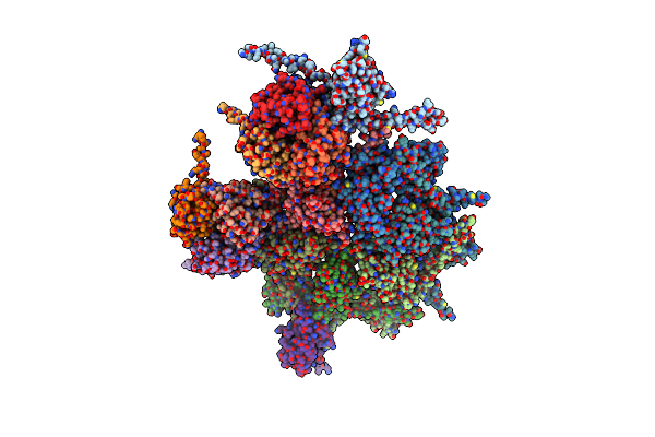
The Composite Cryo-Em Structure Of The Portal Vertex Of Bacteriophage Fcwl1
Organism: Escherichia phage fcwl1
Method: ELECTRON MICROSCOPY Release Date: 2025-01-22 Classification: VIRAL PROTEIN |
 |
Organism: Saccharomyces cerevisiae s288c
Method: ELECTRON MICROSCOPY Release Date: 2024-11-27 Classification: TRANSFERASE Ligands: 6PL, A1D5V, 7PO |
 |
Organism: Saccharomyces cerevisiae s288c
Method: ELECTRON MICROSCOPY Release Date: 2024-11-27 Classification: TRANSFERASE Ligands: 6PL |
 |
Organism: Saccharomyces cerevisiae (strain atcc 204508 / s288c)
Method: ELECTRON MICROSCOPY Release Date: 2024-11-27 Classification: TRANSFERASE Ligands: 6PL, A1D74 |
 |
Organism: Rabbit hemorrhagic disease virus 2
Method: ELECTRON MICROSCOPY Release Date: 2024-10-16 Classification: VIRUS |
 |
Cryo-Em Structure Of A T=1 Vlp Of Rhdv Gi.2 With N-Terminal 1-37 Residues Truncated
Organism: Rabbit hemorrhagic disease virus 2
Method: ELECTRON MICROSCOPY Release Date: 2024-10-16 Classification: VIRUS LIKE PARTICLE |
 |
Organism: Rabbit hemorrhagic disease virus 2
Method: ELECTRON MICROSCOPY Release Date: 2024-10-16 Classification: VIRAL PROTEIN |
 |
Organism: Rabbit hemorrhagic disease virus 2
Method: ELECTRON MICROSCOPY Release Date: 2024-10-16 Classification: VIRUS LIKE PARTICLE |
 |
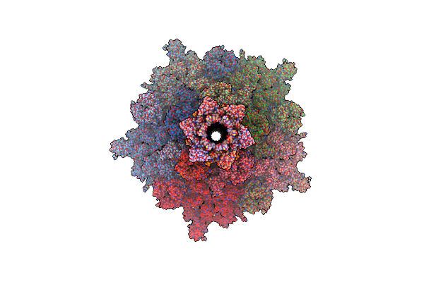
Near-Atomic Structure Of Icosahedrally Averaged Jumbo Bacteriophage Phikz Capsid
Organism: Phikzvirus phikz
Method: ELECTRON MICROSCOPY Release Date: 2024-08-14 Classification: VIRUS |