
Search Count: 142
 |
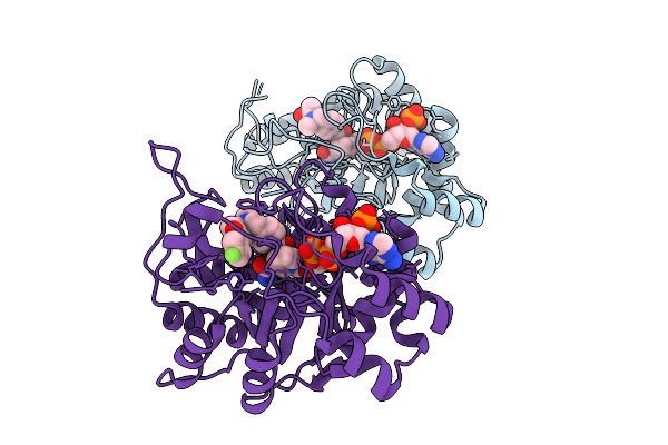
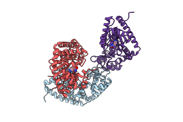
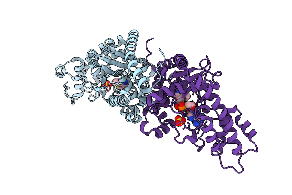
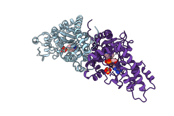
Organism: Escherichia coli k-12
Method: X-RAY DIFFRACTION Release Date: 2025-12-10 Classification: LIGASE Ligands: ATP, PEG, KAA |
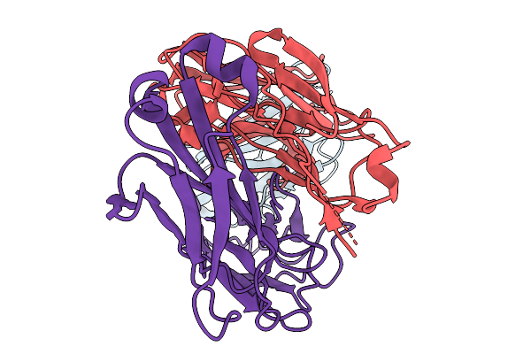 |
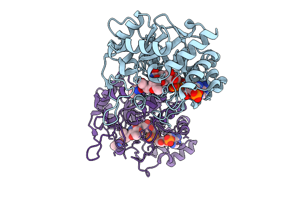
Structure Of Nectin-4 D1 Domain In Complex With The Fab Fragment Of 9Mw2821 Mab
Organism: Mus musculus, Homo sapiens
Method: ELECTRON MICROSCOPY Release Date: 2025-11-19 Classification: IMMUNE SYSTEM |
 |
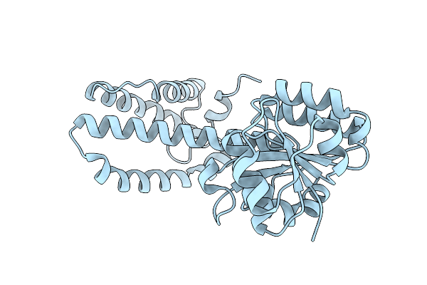
Organism: Micromonospora echinaurantiaca
Method: X-RAY DIFFRACTION Release Date: 2025-11-05 Classification: OXIDOREDUCTASE Ligands: NDP |
 |
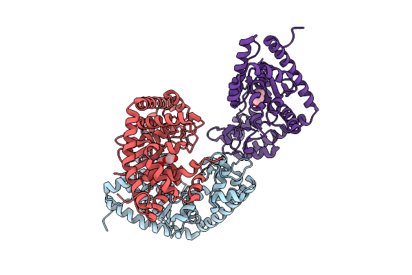
Crystal Structure Of Aldo-Keto Reductase 1C3 Complexed With Compound S30-1023
Organism: Homo sapiens
Method: X-RAY DIFFRACTION Release Date: 2025-10-08 Classification: OXIDOREDUCTASE Ligands: NAP, A1EC0 |
 |
Crystal Structure Of Aldo-Keto Reductase 1C3 Complexed With Compound S30-1023X
Organism: Homo sapiens
Method: X-RAY DIFFRACTION Release Date: 2025-10-08 Classification: OXIDOREDUCTASE Ligands: NAP, A1EC3, SO4 |
 |
Crystal Structure Of Aldo-Keto Reductase 1C3 Complexed With Compound S30-1045
Organism: Homo sapiens
Method: X-RAY DIFFRACTION Release Date: 2025-10-08 Classification: OXIDOREDUCTASE Ligands: NAP, A1EC4 |
 |
Organism: Homo sapiens
Method: X-RAY DIFFRACTION Release Date: 2025-10-01 Classification: OXIDOREDUCTASE Ligands: A1EC7, NAP |
 |
Crystal Structure Of Aldo-Keto Reductase 1C3 Complexed With Compound S30-1018
Organism: Homo sapiens
Method: X-RAY DIFFRACTION Release Date: 2025-10-01 Classification: OXIDOREDUCTASE Ligands: NAP, A1ECZ |
 |
Organism: Micromonospora echinaurantiaca
Method: X-RAY DIFFRACTION Release Date: 2025-07-30 Classification: OXIDOREDUCTASE Ligands: NAP |
 |
Organism: Micromonospora echinaurantiaca
Method: X-RAY DIFFRACTION Release Date: 2025-07-30 Classification: OXIDOREDUCTASE |
 |
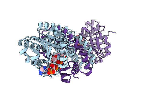
Crystal Structure Of Tryptophanyl-Trna Synthetase From Staphylococcus Aureus
Organism: Staphylococcus aureus
Method: X-RAY DIFFRACTION Release Date: 2025-07-23 Classification: LIGASE Ligands: PEG |
 |
Crystal Structure Of S. Aureus Tryptophanyl-Trna Synthetase Complexed With Tryptophan
Organism: Staphylococcus aureus
Method: X-RAY DIFFRACTION Release Date: 2025-07-23 Classification: LIGASE Ligands: TRP |
 |
Crystal Structure Of S. Aureus Tryptophanyl-Trna Synthetase Complexed With Chuangxinmycin
Organism: Staphylococcus aureus
Method: X-RAY DIFFRACTION Release Date: 2025-07-23 Classification: LIGASE Ligands: 9E0 |
 |
Crystal Structure Of S. Aureus Tryptophanyl-Trna Synthetase Complexed With 3-Demethylchuangxinmycin
Organism: Staphylococcus aureus
Method: X-RAY DIFFRACTION Release Date: 2025-07-23 Classification: LIGASE Ligands: A1EHM |
 |
Crystal Structure Of S. Aureus Tryptophanyl-Trna Synthetase Complexed With 3-Methylchuangxinmycin
Organism: Staphylococcus aureus
Method: X-RAY DIFFRACTION Release Date: 2025-07-23 Classification: LIGASE Ligands: A1EHN |
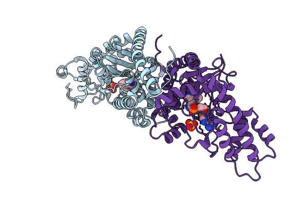 |
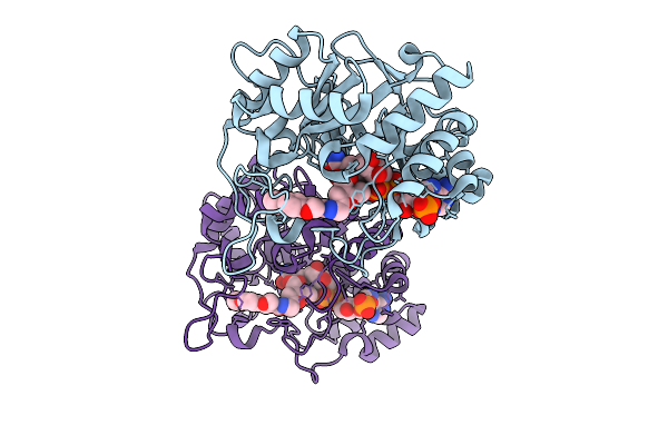
Crystal Structure Of E. Coli Tryptophanyl-Trna Synthetase Complexed With Chuangxinmycin And Tryptophanyl-5'-Amp
Organism: Escherichia coli
Method: X-RAY DIFFRACTION Release Date: 2025-07-23 Classification: LIGASE Ligands: 9E0, SO4, TYM |
 |
Crystal Structure Of E. Coli Tryptophanyl-Trna Synthetase Complexed With 3-Demethylchuangxinmycin And Tryptophanyl-5'-Amp
Organism: Escherichia coli
Method: X-RAY DIFFRACTION Release Date: 2025-07-23 Classification: LIGASE Ligands: A1EHM, SO4, TYM |
 |
Crystal Structure Of E. Coli Tryptophanyl-Trna Synthetase Complexed With 3-Methylchuangxinmycin And Tryptophanyl-5'-Amp
Organism: Escherichia coli
Method: X-RAY DIFFRACTION Release Date: 2025-07-23 Classification: LIGASE Ligands: A1EHN, SO4, TYM |
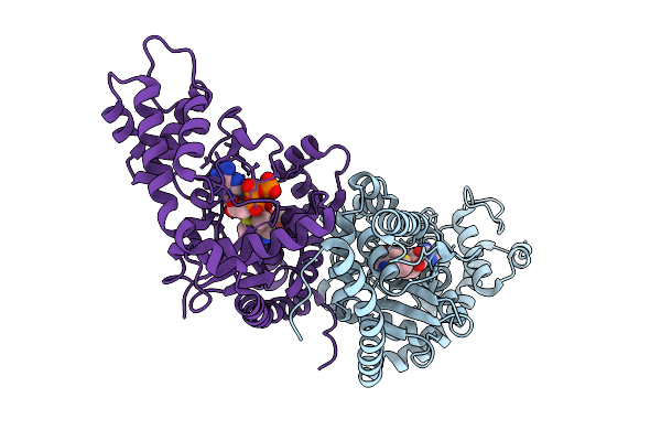 |
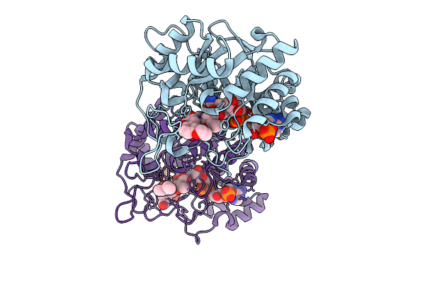
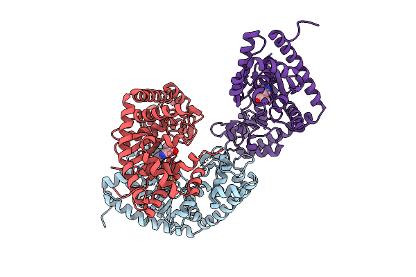
Crystal Structure Of E. Coli Tryptophanyl-Trna Synthetase Complexed With Chuangxinmycin And Atp In Closed-Closed State
Organism: Escherichia coli
Method: X-RAY DIFFRACTION Release Date: 2025-07-23 Classification: LIGASE Ligands: 9E0, ATP, MG, GOL |
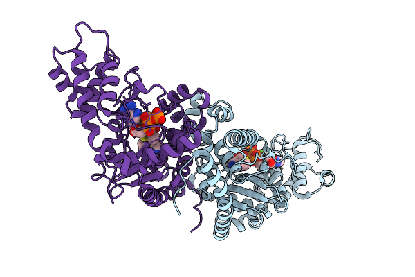 |
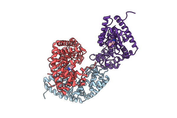
Crystal Structure Of E. Coli Tryptophanyl-Trna Synthetase Complexed With Chuangxinmycin And Atp In Open-Closed State
Organism: Escherichia coli
Method: X-RAY DIFFRACTION Release Date: 2025-07-23 Classification: LIGASE Ligands: 9E0, ATP, MG |