
Search Count: 3
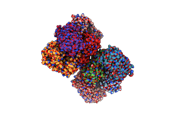 |
Crystal Structure Of The Glycosyltransferase Babsha Bound With Udp And L-Malate
Organism: Bacillus anthracis
Method: X-RAY DIFFRACTION Resolution:3.31 Å Release Date: 2010-09-29 Classification: TRANSFERASE Ligands: UDP, GOL, MLT, MG |
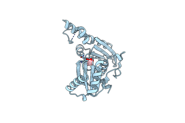 |
Structure Of The Type Iii Pantothenate Kinase (Coax) From Bacillus Anthracis
Organism: Bacillus anthracis str.
Method: X-RAY DIFFRACTION Resolution:2.00 Å Release Date: 2007-03-20 Classification: BIOSYNTHETIC PROTEIN Ligands: EDO |
 |
The Crystal Structure Of 1D-Myo-Inositol 2-Acetamido-2-Deoxy-Alpha-D-Glucopyranoside Deacetylase (Mshb)
Organism: Mycobacterium tuberculosis
Method: X-RAY DIFFRACTION Resolution:1.70 Å Release Date: 2003-12-02 Classification: HYDROLASE Ligands: ZN, PE4 |