
Search Count: 13
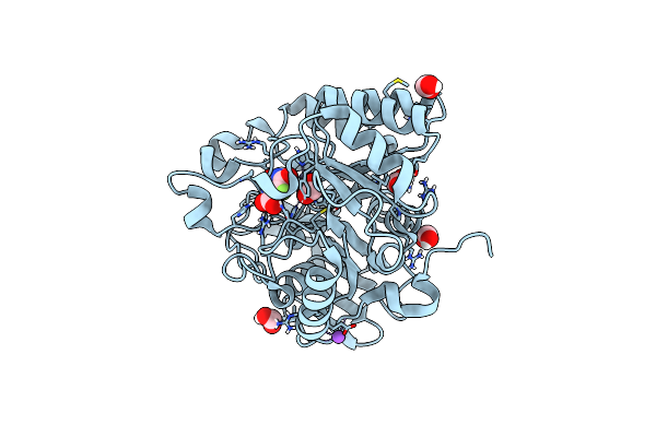 |
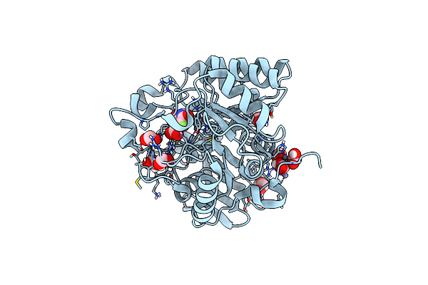
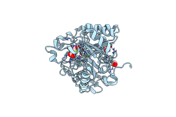
Mutant K1556T Of The Dihydroorotase Domain Of Human Cad Protein Bound To The Inhibitor Fluoroorotate
Organism: Homo sapiens
Method: X-RAY DIFFRACTION Resolution:1.46 Å Release Date: 2023-11-01 Classification: HYDROLASE Ligands: FMT, FOT, ZN, NA |
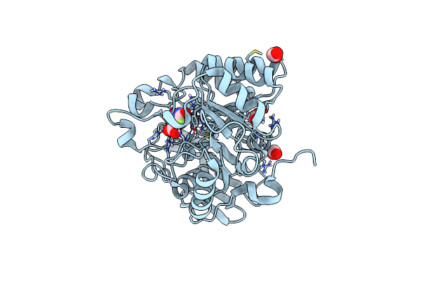 |
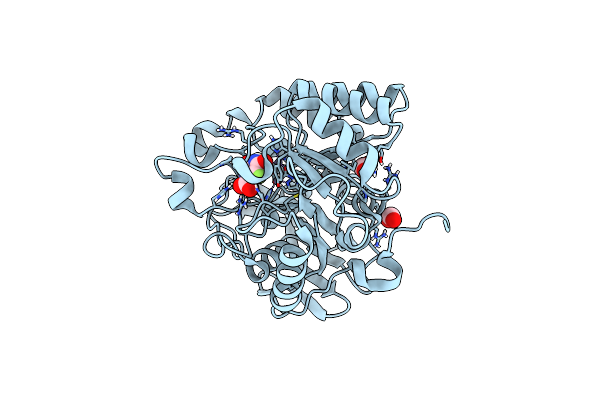
Mutant R1617Q Of The Dihydroorotase Domain Of Human Cad Protein Bound To The Inhibitor Fluoroorotate
Organism: Homo sapiens
Method: X-RAY DIFFRACTION Resolution:1.50 Å Release Date: 2023-11-01 Classification: HYDROLASE Ligands: FOT, FMT, ZN |
 |
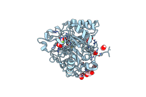
Mutant R1722W Of The Dihydroorotase Domain Of Human Cad Protein Bound To The Inhibitor Fluoorotate
Organism: Homo sapiens
Method: X-RAY DIFFRACTION Resolution:1.69 Å Release Date: 2023-11-01 Classification: HYDROLASE Ligands: FOT, GOL, FMT, ZN |
 |
Mutant R1789Q Of The Dihydroorotase Domain Of Human Cad Protein Bound To The Inhibitor Fluoorotate
Organism: Homo sapiens
Method: X-RAY DIFFRACTION Resolution:1.71 Å Release Date: 2023-11-01 Classification: HYDROLASE Ligands: FOT, ZN, FMT, GOL |
 |
Mutant K1482M Of The Dihydroorotase Domain Of Human Cad Protein Bound To The Inhibitor Fluoorotate
Organism: Homo sapiens
Method: X-RAY DIFFRACTION Resolution:1.67 Å Release Date: 2023-11-01 Classification: HYDROLASE Ligands: FOT, ZN, FMT |
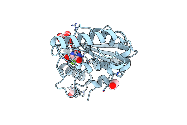 |
Crystal Structure Of Mycobacterium Tuberculosis Uracil-Dna Glycosylase In Complex With 5-Fluoroorotic Acid And Citric Acid, Form I
Organism: Mycobacterium tuberculosis h37rv
Method: X-RAY DIFFRACTION Resolution:2.00 Å Release Date: 2023-07-12 Classification: HYDROLASE/INHIBITOR Ligands: FOT, CIT, EDO |
 |
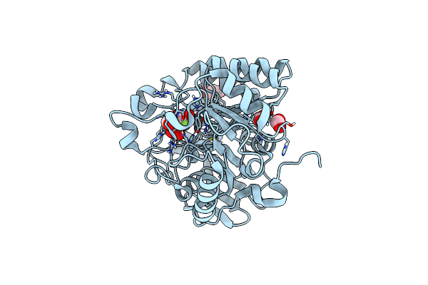
Crystal Structure Of Dihydroorotase In Complex With 5-Fluoroorotic Acid From Saccharomyces Cerevisiae
Organism: Saccharomyces cerevisiae (strain atcc 204508 / s288c)
Method: X-RAY DIFFRACTION Resolution:2.50 Å Release Date: 2021-06-09 Classification: HYDROLASE Ligands: ZN, FOT |
 |
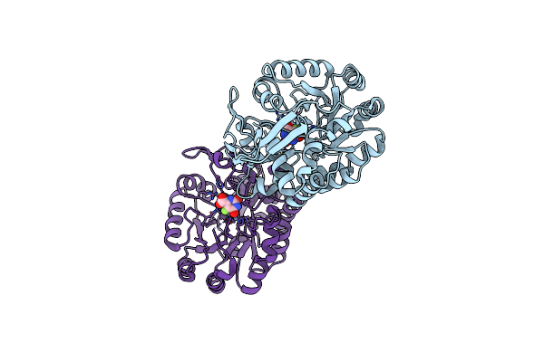
Hybrid Dihydroorotase Domain Of Human Cad With E. Coli Flexible Loop, Bound To Fluoroorotate
Organism: Homo sapiens
Method: X-RAY DIFFRACTION Resolution:1.83 Å Release Date: 2018-10-24 Classification: BIOSYNTHETIC PROTEIN Ligands: ZN, FOT, FMT |
 |
Crystal Structure Of The Dihydroorotase Domain Of Human Cad Bound To The Inhibitor Fluoroorotate At Ph 6.0
Organism: Homo sapiens
Method: X-RAY DIFFRACTION Resolution:1.55 Å Release Date: 2014-02-05 Classification: HYDROLASE Ligands: ZN, FMT, FOT |
 |
Crystal Structure Of The Dihydroorotase Domain Of Human Cad Bound To The Inhibitor Fluoroorotate At Ph 7.0
Organism: Homo sapiens
Method: X-RAY DIFFRACTION Resolution:1.62 Å Release Date: 2014-01-08 Classification: HYDROLASE Ligands: ZN, FMT, FOT |
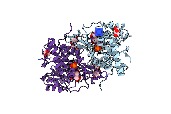 |
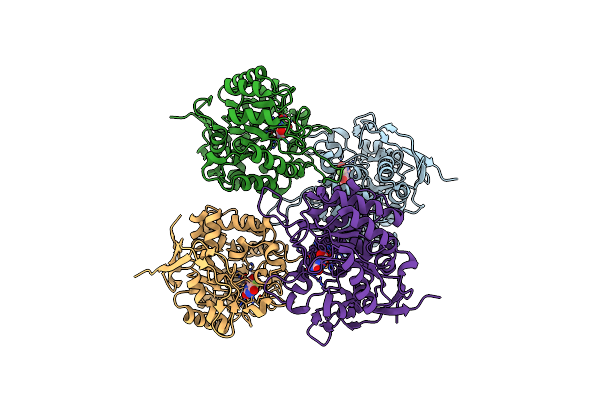
Structure Of Trypanosoma Cruzi Dihydroorotate Dehydrogenase In Complex With 5-Halogenated Orotate Derivatives
Organism: Trypanosoma cruzi
Method: X-RAY DIFFRACTION Resolution:1.42 Å Release Date: 2013-11-13 Classification: OXIDOREDUCTASE Ligands: FMN, NCO, FOT, GOL |
 |
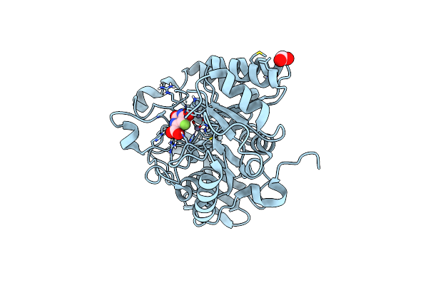
The Crystal Structure Of E. Coli Dihydroorotase Complexed With 5-Fluoroorotic Acid
Organism: Escherichia coli
Method: X-RAY DIFFRACTION Resolution:2.20 Å Release Date: 2007-07-03 Classification: HYDROLASE Ligands: ZN, FOT |
 |
The Crystal Structure Of The T109S Mutant Of E. Coli Dihydroorotase Complexed With An Inhibitor 5-Fluoroorotate
Organism: Escherichia coli
Method: X-RAY DIFFRACTION Resolution:2.70 Å Release Date: 2007-03-13 Classification: HYDROLASE Ligands: ZN, FOT |