
Search Count: 27
All
Selected
 |
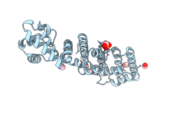
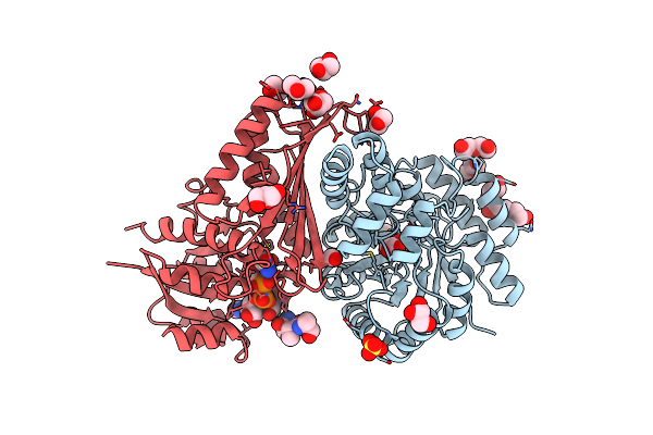
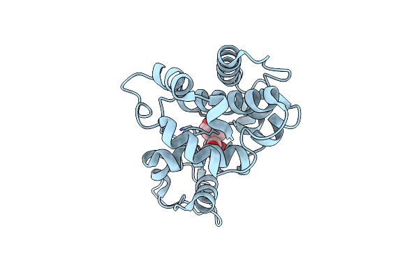
Crystal Structure Of Sr2+-Bound Rtx Domain Block V Of Adenylate Cyclase Toxin From Bordetella Pertussis
Organism: Bordetella pertussis
Method: X-RAY DIFFRACTION Resolution:1.50 Å Release Date: 2026-01-07 Classification: TOXIN Ligands: SR, CL, NA, FOR, GOL, TRS |
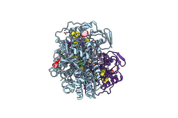 |
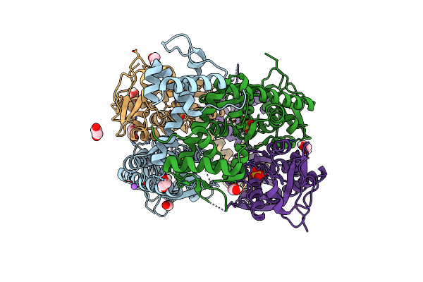
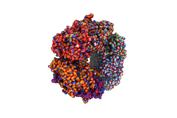
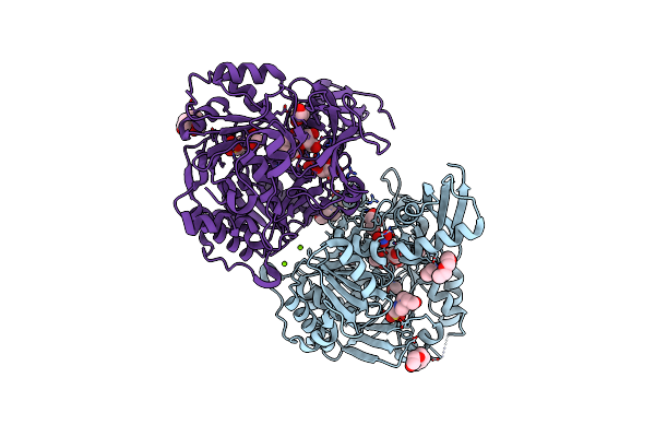
Formaldehyde-Inhibited [Fefe]-Hydrogenase Cpi From Clostridium Pasteurianum, Variant C299D
Organism: Clostridium pasteurianum
Method: X-RAY DIFFRACTION Resolution:1.38 Å Release Date: 2023-11-29 Classification: OXIDOREDUCTASE Ligands: 402, SF4, FES, 74C, GOL, FOR, MG, CL |
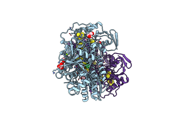 |
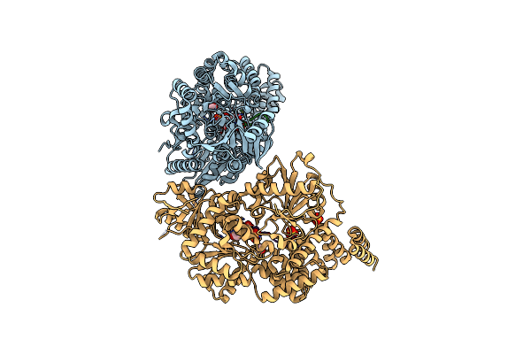
Formaldehyde-Inhibited [Fefe]-Hydrogenase I From Clostridium Pasteurianum (Cpi)
Organism: Clostridium pasteurianum
Method: X-RAY DIFFRACTION Resolution:1.53 Å Release Date: 2023-11-29 Classification: OXIDOREDUCTASE Ligands: 402, SF4, FES, FOR, GOL, CL, MG |
 |
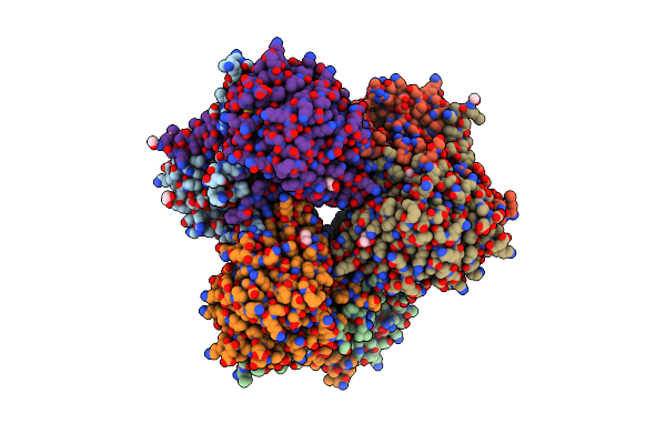
Organism: Aspergillus fumigatus af293
Method: X-RAY DIFFRACTION Resolution:1.06 Å Release Date: 2023-11-01 Classification: HYDROLASE Ligands: IMD, EDO, FOR, NA |
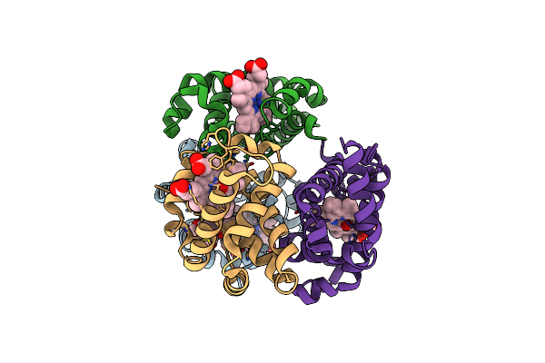 |
Crystal Structure Of Carbonmonoxy Hemoglobin S (Liganded Sickle Cell Hemoglobin) Complexed With Gbt021601
Organism: Homo sapiens
Method: X-RAY DIFFRACTION Resolution:2.05 Å Release Date: 2023-04-05 Classification: OXYGEN TRANSPORT Ligands: HEM, FOR, OHF |
 |
Organism: Candida parapsilosis, Streptomyces albus
Method: X-RAY DIFFRACTION Resolution:1.35 Å Release Date: 2021-04-21 Classification: ANTIBIOTIC Ligands: MES, PEG, FOR, PGE |
 |
Organism: Candida parapsilosis, Streptomyces albus
Method: X-RAY DIFFRACTION Resolution:1.80 Å Release Date: 2021-04-21 Classification: ANTIBIOTIC Ligands: 1PE, PEG, PGE, MES, NA, FOR |
 |
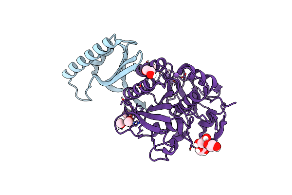
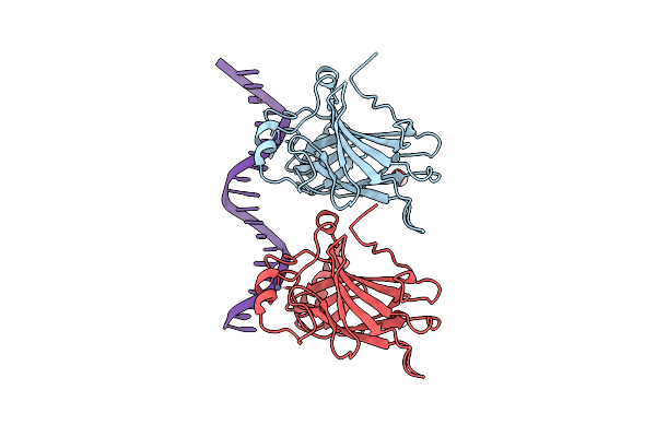
Structure Of Bacillus Subtilis Hxlr, Wild Type In Complex With Formaldehyde And Dna
Organism: Bacillus subtilis (strain 168), Bacillus subtilis subsp. subtilis str. 168
Method: X-RAY DIFFRACTION Resolution:2.90 Å Release Date: 2021-02-03 Classification: DNA BINDING PROTEIN Ligands: FOR, PGE, MG, PE8, PEG |
 |
Organism: Corynebacterium glutamicum
Method: X-RAY DIFFRACTION Resolution:1.99 Å Release Date: 2018-12-05 Classification: TRANSCRIPTION Ligands: EDO, FMT, SO4, PEG, NA, FOR |
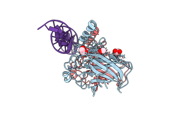 |
New Insights Into The Role Of Dna Shape On Its Recognition By P53 Proteins (Complex P53Dbd-Lhg1)
Organism: Homo sapiens, Synthetic construct
Method: X-RAY DIFFRACTION Resolution:1.45 Å Release Date: 2018-06-13 Classification: TRANSCRIPTION Ligands: ZN, EDO, FOR |
 |
New Insights Into The Role Of Dna Shape On Its Recognition By P53 Proteins (Complex P53Dbd-Lwc2)
Organism: Homo sapiens, Synthetic construct
Method: X-RAY DIFFRACTION Resolution:1.90 Å Release Date: 2018-06-13 Classification: TRANSCRIPTION Ligands: ZN, FOR |
 |
The Asymmetric Tetrameric Structure Of The Formaldehyde Sensing Transcriptional Repressor Frmr From Escherichia Coli
Organism: Escherichia coli o157:h7
Method: X-RAY DIFFRACTION Resolution:2.70 Å Release Date: 2016-12-21 Classification: TRANSCRIPTION Ligands: FOR |
 |
Crystal Structure Of 7,8-Diaminopelargonic Acid Synthase (Bioa) From Mycobacterium Tuberculosis, Complexed With A Thiazole Benzamide Inhibitor
Organism: Mycobacterium tuberculosis
Method: X-RAY DIFFRACTION Resolution:1.70 Å Release Date: 2015-07-08 Classification: transferase/transferase inhibitor Ligands: PLP, 3VS, EDO, FOR, SO4, MG |
 |
Organism: Chlamydia pneumoniae
Method: X-RAY DIFFRACTION Resolution:1.20 Å Release Date: 2014-07-30 Classification: TRANSPORT PROTEIN Ligands: FOR, SO4, CL, DMN |
 |
Crystal And Solution Structures Of The Bifunctional Enzyme (Aldolase/Aldehyde Dehydrogenase) From Thermomonospora Curvata, Reveal A Cofactor-Binding Domain Motion During Nad+ And Coa Accommodation Whithin The Shared Cofactor-Binding Site
Organism: Thermomonospora curvata
Method: X-RAY DIFFRACTION Resolution:1.55 Å Release Date: 2013-09-04 Classification: OXIDOREDUCTASE Ligands: SO4, PYR, CL, MG, GOL, PEG, NAD, FOR |
 |
Crystal Structure Of Hansenula Polymorpha Copper Amine Oxidase-1 Reduced By Methylamine At Ph 7.0
Organism: Ogataea angusta
Method: X-RAY DIFFRACTION Resolution:2.10 Å Release Date: 2013-08-21 Classification: OXIDOREDUCTASE Ligands: CU, PEO, FOR, GOL, PO4 |
 |
A Neutral Diphosphate Mimic Crosslinks The Active Site Of Human O-Glcnac Transferase
Organism: Homo sapiens
Method: X-RAY DIFFRACTION Resolution:1.88 Å Release Date: 2011-11-16 Classification: TRANSFERASE/TRANSFERASE INHIBITOR Ligands: UDP, FOR, SO4 |
 |
Organism: Escherichia coli
Method: X-RAY DIFFRACTION Resolution:2.59 Å Release Date: 2010-04-14 Classification: HYDROLASE Ligands: GOL, FOR |
 |
Organism: Picea abies
Method: X-RAY DIFFRACTION Resolution:1.55 Å Release Date: 2009-08-11 Classification: HYDROLASE Ligands: MXE, ACT, FOR |
 |
2.1 A Structure Of Acyl-Adenylate Synthetase From Methanosarcina Acetivorans Containing A Link Between Lys256 And Cys298
Organism: Methanosarcina acetivorans
Method: X-RAY DIFFRACTION Resolution:2.10 Å Release Date: 2009-07-07 Classification: LIGASE Ligands: FOR, PGE, 1PE, EPE, MG, NO3, GOL |