
Search Count: 4
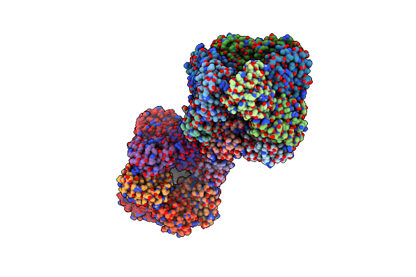 |
Crystal Structure Of 3-Dehydroquinate Dehydratase From Mycobacterium Tuberculosis In Complex With Inhibitor 2
Organism: Mycobacterium tuberculosis
Method: X-RAY DIFFRACTION Resolution:2.00 Å Release Date: 2011-05-11 Classification: LYASE/LYASE INHIBITOR Ligands: FA1, GOL |
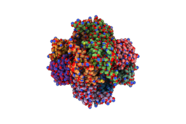 |
Organism: Helicobacter pylori
Method: X-RAY DIFFRACTION Resolution:3.10 Å Release Date: 2006-02-22 Classification: LYASE Ligands: FA1 |
 |
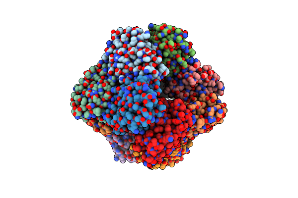
Type Ii Dehydroquinase From Mycobacterium Tuberculosis Complexed With 2,3-Anhydro-Quinic Acid
Organism: Mycobacterium tuberculosis
Method: X-RAY DIFFRACTION Resolution:2.10 Å Release Date: 2003-10-23 Classification: DEHYDRATASE Ligands: FA1, PO4, CL, GOL |
 |
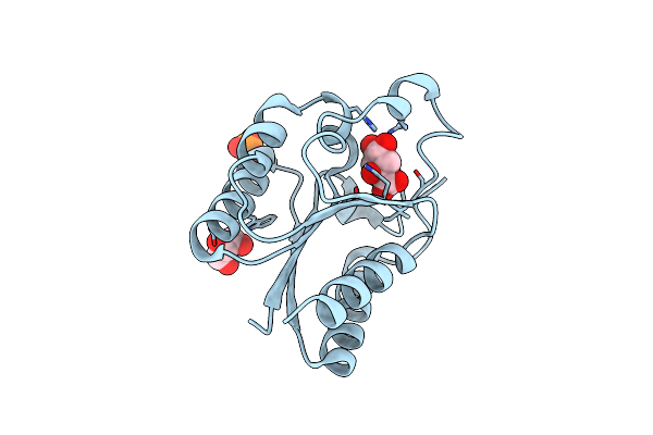
Crystal Structure Of Type Ii Dehydroquinase From Streptomyces Coelicolor Complexed With 2,3-Anhydro-Quinic Acid
Organism: Streptomyces coelicolor
Method: X-RAY DIFFRACTION Resolution:1.80 Å Release Date: 2002-04-12 Classification: LYASE Ligands: FA1, GOL, TLA, TRS |