
Search Count: 15
 |
Organism: Homo sapiens, Severe acute respiratory syndrome coronavirus 2
Method: X-RAY DIFFRACTION Resolution:3.30 Å Release Date: 2024-05-15 Classification: VIRAL PROTEIN/IMMUNE SYSTEM |
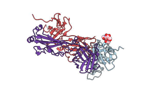 |
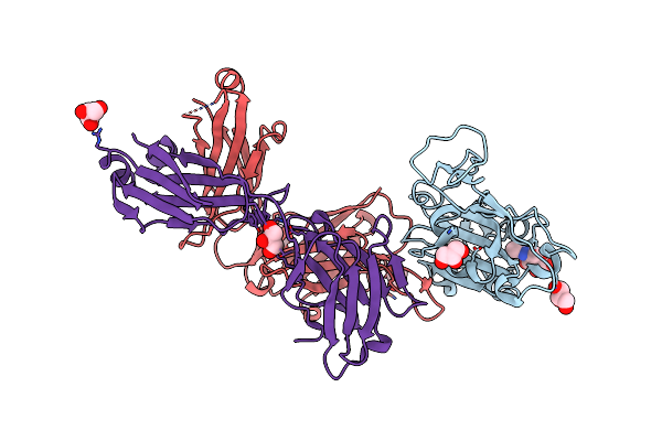
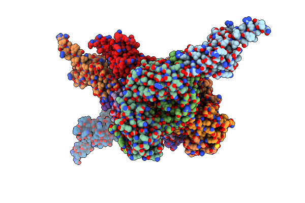
Crystal Structure Of Sars-Cov-2 Receptor Binding Domain In Complex With Sars-Cov-2 Reactive Human Antibody Cr3022
Organism: Homo sapiens, Severe acute respiratory syndrome coronavirus 2
Method: X-RAY DIFFRACTION Resolution:4.22 Å Release Date: 2023-12-13 Classification: VIRAL PROTEIN/IMMUNE SYSTEM |
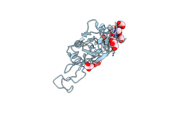 |
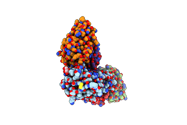
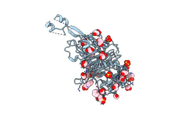
Organism: Severe acute respiratory syndrome coronavirus 2
Method: X-RAY DIFFRACTION Resolution:1.95 Å Release Date: 2023-12-13 Classification: VIRAL PROTEIN Ligands: NAG, FUC, GOL |
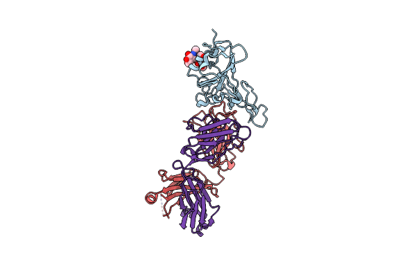 |
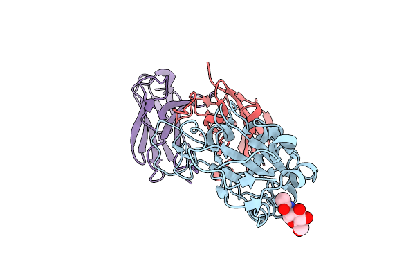
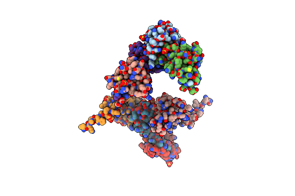
Crystal Structure Of Antibody Wrair-2123 In Complex With Sars-Cov-2 Receptor Binding Domain
Organism: Severe acute respiratory syndrome coronavirus 2, Homo sapiens
Method: X-RAY DIFFRACTION Resolution:3.50 Å Release Date: 2023-12-13 Classification: VIRAL PROTEIN/IMMUNE SYSTEM |
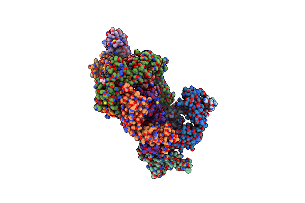 |
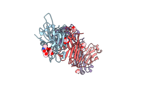
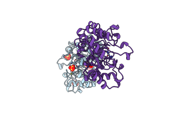
Crystal Structure Of Antibody Wrair-2134 In Complex With Sars-Cov-2 Receptor Binding Domain
Organism: Homo sapiens, Severe acute respiratory syndrome coronavirus 2
Method: X-RAY DIFFRACTION Resolution:3.16 Å Release Date: 2023-06-28 Classification: VIRAL PROTEIN/IMMUNE SYSTEM Ligands: NAG |
 |
Crystal Structure Of Antibody Ab246 In Complex With Sars-Cov-2 Receptor Binding Domain
Organism: Severe acute respiratory syndrome coronavirus 2, Homo sapiens
Method: X-RAY DIFFRACTION Resolution:2.29 Å Release Date: 2023-03-15 Classification: VIRAL PROTEIN/IMMUNE SYSTEM Ligands: NAG, GOL, CL |
 |
Organism: Homo sapiens, Severe acute respiratory syndrome coronavirus 2
Method: ELECTRON MICROSCOPY Release Date: 2022-10-19 Classification: VIRAL PROTEIN/IMMUNE SYSTEM Ligands: NAG |
 |
Organism: Homo sapiens, Severe acute respiratory syndrome coronavirus 2
Method: ELECTRON MICROSCOPY Release Date: 2022-10-19 Classification: VIRAL PROTEIN/IMMUNE SYSTEM Ligands: NAG |
 |
Crystal Structure Of Sars-Cov-2 Spike Rbd In Complex With Human Monoclonal Antibody Azd8895
Organism: Homo sapiens, Severe acute respiratory syndrome coronavirus 2
Method: X-RAY DIFFRACTION Resolution:2.50 Å Release Date: 2021-09-01 Classification: VIRAL PROTEIN/IMMUNE SYSTEM Ligands: GOL, NAG |
 |
Crystal Structure Of Sars-Cov-2 Spike Rbd In Complex With Human Monoclonal Antibodies Azd8895 And Azd1061
Organism: Homo sapiens, Severe acute respiratory syndrome coronavirus 2
Method: X-RAY DIFFRACTION Resolution:3.00 Å Release Date: 2021-09-01 Classification: VIRAL PROTEIN/IMMUNE SYSTEM Ligands: PO4, NAG |
 |
Organism: Pseudomonas aeruginosa, Homo sapiens
Method: X-RAY DIFFRACTION Resolution:2.78 Å Release Date: 2018-08-01 Classification: IMMUNE SYSTEM |
 |
Organism: Escherichia coli
Method: X-RAY DIFFRACTION Resolution:2.50 Å Release Date: 2014-05-07 Classification: TRANSFERASE Ligands: SO4, CXS, GOL, EDO, CL |
 |
Crystal Structure Of E. Coli Penicillin Binding Protein 3, Domain V88- S165
Organism: Escherichia coli
Method: X-RAY DIFFRACTION Resolution:2.10 Å Release Date: 2014-05-07 Classification: TRANSFERASE Ligands: SO4, TRS |
 |
Organism: Escherichia coli
Method: X-RAY DIFFRACTION Resolution:1.70 Å Release Date: 1999-06-08 Classification: PHOSPHOTRIESTERASE Ligands: ZN, SO4, MPD, GOL |
 |
Organism: Tachyglossus aculeatus
Method: X-RAY DIFFRACTION Resolution:1.90 Å Release Date: 1997-04-21 Classification: LYSOZYME Ligands: CA |