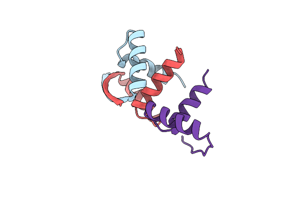
Search Count: 17
 |
Organism: Homo sapiens
Method: ELECTRON MICROSCOPY Release Date: 2025-04-02 Classification: VIRUS LIKE PARTICLE |
 |
Organism: Homo sapiens
Method: ELECTRON MICROSCOPY Release Date: 2025-03-26 Classification: VIRUS LIKE PARTICLE |
 |
Organism: Human picobirnavirus
Method: ELECTRON MICROSCOPY Release Date: 2020-09-23 Classification: VIRUS LIKE PARTICLE |
 |
Organism: Human picobirnavirus
Method: ELECTRON MICROSCOPY Release Date: 2020-09-23 Classification: VIRUS LIKE PARTICLE |
 |
Organism: Human picobirnavirus (strain human/thailand/hy005102/-)
Method: ELECTRON MICROSCOPY Release Date: 2020-09-16 Classification: VIRUS LIKE PARTICLE |
 |
Organism: Streptococcus agalactiae, Plasmid pmv158
Method: X-RAY DIFFRACTION Resolution:2.00 Å Release Date: 2017-04-12 Classification: DNA BINDING PROTEIN Ligands: CL, MG, GOL, NA |
 |
Mobm Relaxase Domain (Mobv; Mob_Pre) Bound To Plasmid Pmv158 Orit Dna (22Nt). Mn-Bound Crystal Structure At Ph 4.6
Organism: Streptococcus agalactiae
Method: X-RAY DIFFRACTION Resolution:1.90 Å Release Date: 2014-09-24 Classification: DNA BINDING PROTEIN/DNA Ligands: MN, GOL |
 |
Mobm Relaxase Domain (Mobv; Mob_Pre) Bound To Plasmid Pmv158 Orit Dna (22Nt). Mn-Bound Crystal Structure At Ph 5.5
Organism: Streptococcus agalactiae
Method: X-RAY DIFFRACTION Resolution:2.17 Å Release Date: 2014-09-24 Classification: DNA BINDING PROTEIN/DNA Ligands: MN, ACT |
 |
Mobm Relaxase Domain (Mobv; Mob_Pre) Bound To Plasmid Pmv158 Orit Dna (22Nt+3'Phosphate). Mn-Bound Crystal Structure At Ph 4.6
Organism: Streptococcus agalactiae
Method: X-RAY DIFFRACTION Resolution:2.37 Å Release Date: 2014-09-24 Classification: DNA BINDING PROTEIN/DNA Ligands: MN, NA |
 |
Mobm Relaxase Domain (Mobv; Mob_Pre) Bound To Plasmid Pmv158 Orit Dna (22Nt+3'Thiophosphate). Mn-Bound Crystal Structure At Ph 6.8
Organism: Streptococcus agalactiae, Synthetic dna
Method: X-RAY DIFFRACTION Resolution:2.20 Å Release Date: 2014-09-24 Classification: DNA BINDING PROTEIN/DNA Ligands: MN, CL |
 |
Mobm Relaxase Domain (Mobv; Mob_Pre) Bound To Plasmid Pmv158 Orit Dna (23Nt). Mn-Bound Crystal Structure At Ph 6.5
Organism: Streptococcus agalactiae
Method: X-RAY DIFFRACTION Resolution:3.10 Å Release Date: 2014-09-24 Classification: DNA BINDING PROTEIN/DNA Ligands: MN, MG, CL, NA |
 |
Crystal Structure Of The Replication Initiator Protein Encoded On Plasmid Pmv158 (Repb), Trigonal Form, To 2.7 Ang Resolution
Organism: Streptococcus agalactiae
Method: X-RAY DIFFRACTION Resolution:2.70 Å Release Date: 2009-06-30 Classification: REPLICATION Ligands: MN, CL, MG |
 |
Crystal Structure Of The Replication Initiator Protein Encoded On Plasmid Pmv158 (Repb), Tetragonal Form, To 3.6 Ang Resolution
Organism: Streptococcus agalactiae
Method: X-RAY DIFFRACTION Resolution:3.60 Å Release Date: 2009-06-30 Classification: REPLICATION Ligands: MN |
 |
Organism: Streptococcus agalactiae
Method: X-RAY DIFFRACTION Resolution:2.95 Å Release Date: 2001-07-05 Classification: GENE REGULATION/DNA |
 |
Organism: Penicillium simplicissimum
Method: X-RAY DIFFRACTION Resolution:2.75 Å Release Date: 2000-04-12 Classification: FLAVOENZYME Ligands: FAD, FCR |
 |
Organism: Streptococcus agalactiae
Method: X-RAY DIFFRACTION Resolution:2.56 Å Release Date: 1999-11-19 Classification: GENE REGULATION/DNA |
 |
Organism: Streptococcus agalactiae
Method: X-RAY DIFFRACTION Resolution:1.60 Å Release Date: 1999-11-19 Classification: GENE REGULATION Ligands: CL |

