
Search Count: 15
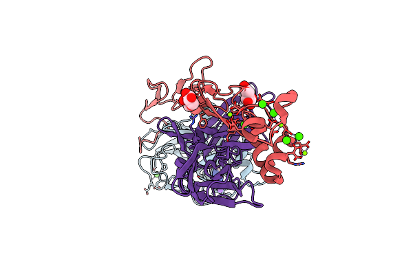 |
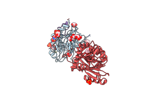
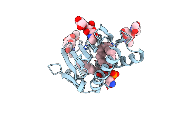
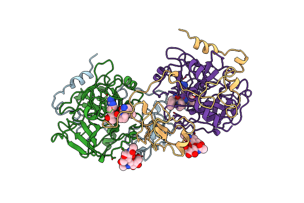
Mg2+ Is Required For Optimal Folding Of The Gamma-Carboxyglutamic Acid (Gla) Domains Of Vitamin K-Dependent Clotting Factors At Physiological Ca2+
Organism: Homo sapiens
Method: X-RAY DIFFRACTION Resolution:1.72 Å Release Date: 2012-08-22 Classification: HYDROLASE/HYDROLASE INHIBITOR Ligands: BGC, FUC, MG, CA, BEN, NA, CL |
 |
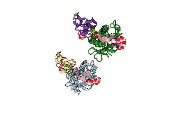
Mg2+ Is Required For Optimal Folding Of The Gamma-Carboxyglutamic Acid (Gla) Domains Of Vitamin K-Dependent Clotting Factors At Physiological Ca2+
Organism: Homo sapiens
Method: X-RAY DIFFRACTION Resolution:2.70 Å Release Date: 2012-08-22 Classification: HYDROLASE/HYDROLASE INHIBITOR Ligands: FUC, CA, BGC, 0GE, CL |
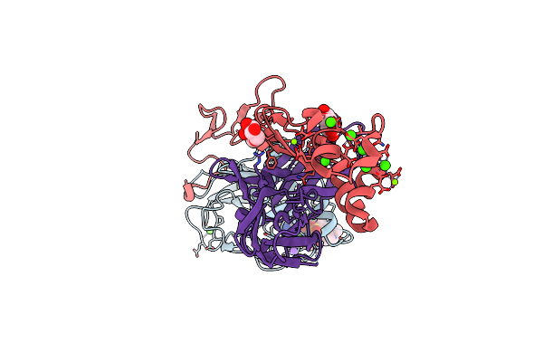 |
Mg2+ Is Required For Optimal Folding Of The Gamma-Carboxyglutamic Acid (Gla) Domains Of Vitamin K-Dependent Clotting Factors At Physiological Ca2+
Organism: Homo sapiens
Method: X-RAY DIFFRACTION Resolution:1.80 Å Release Date: 2012-08-22 Classification: HYDROLASE/HYDROLASE INHIBITOR Ligands: BGC, FUC, MG, CA, 0GE, CL, NA |
 |
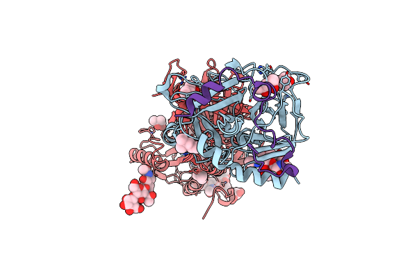
Importance Of Mg2+ In The Ca2+-Dependent Folding Of The Gamma-Carboxyglutamic Acid Domains Of Vitamin K-Dependent Clotting And Anticlotting Proteins
Organism: Homo sapiens
Method: X-RAY DIFFRACTION Resolution:1.60 Å Release Date: 2011-04-06 Classification: BLOOD CLOTTING Ligands: NAG, PTY, MG, CA |
 |
Organism: Homo sapiens
Method: X-RAY DIFFRACTION Resolution:1.60 Å Release Date: 2008-04-22 Classification: HYDROLASE/HYDROLASE INHIBITOR Ligands: SO4, GOL |
 |
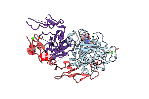
2.5A Crystal Structure Of The Antithrombin-Thrombin-Heparin Ternary Complex
Organism: Homo sapiens
Method: X-RAY DIFFRACTION Resolution:2.50 Å Release Date: 2004-08-17 Classification: HYDROLASE/BLOOD CLOTTING Ligands: MPD, NAG |
 |
Organism: Homo sapiens
Method: X-RAY DIFFRACTION Resolution:2.35 Å Release Date: 2002-08-30 Classification: BLOOD CLOTTING Ligands: CA, NAG |
 |
Organism: Homo sapiens
Method: X-RAY DIFFRACTION Resolution:2.20 Å Release Date: 2002-08-30 Classification: BLOOD CLOTTING |
 |
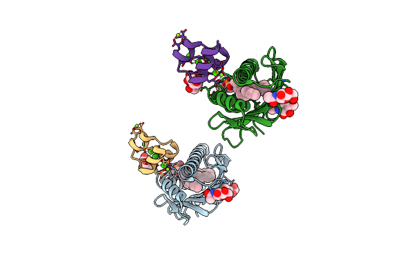
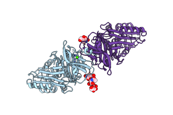
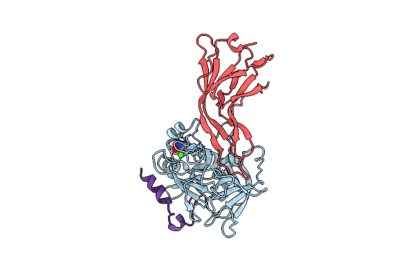
Crystal Structure Of The Endothelial Protein C Receptor And Bound Phospholipid Molecule
Organism: Homo sapiens
Method: X-RAY DIFFRACTION Resolution:2.00 Å Release Date: 2002-06-26 Classification: BLOOD CLOTTING Ligands: PTY |
 |
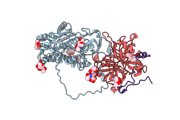
Crystal Structure Of The Endothelial Protein C Receptor With Phospholipid In The Groove In Complex With Gla Domain Of Protein C.
Organism: Homo sapiens
Method: X-RAY DIFFRACTION Resolution:1.60 Å Release Date: 2002-06-19 Classification: BLOOD CLOTTING Ligands: NAG, PTY, CA |
 |
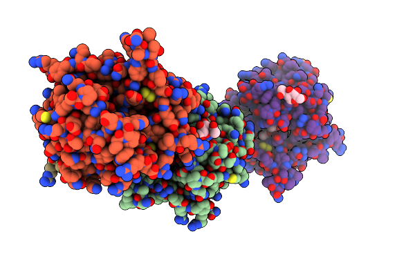
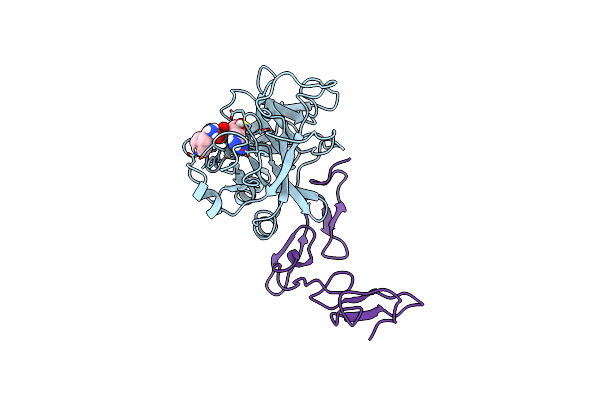
Crystal Structure Of Bovine Thrombin Complex With Protease Inhibitor Ecotin
Organism: Escherichia coli, Bos taurus
Method: X-RAY DIFFRACTION Resolution:2.50 Å Release Date: 2001-09-05 Classification: HYDROLASE Ligands: CA, TRS |
 |
Organism: Homo sapiens
Method: X-RAY DIFFRACTION Resolution:1.80 Å Release Date: 2001-08-10 Classification: BLOOD CLOTTING Ligands: NAG, NA, GOL, ACY |
 |
The X-Ray Crystal Structure Of Ppack-Meizothrombin Desf1: Kringle/Thrombin And Carbohydrate/Kringle/Thrombin Interactions And Location Of The Linker Chain
Organism: Bos taurus
Method: X-RAY DIFFRACTION Resolution:3.20 Å Release Date: 1998-06-17 Classification: HYDROLASE/HYDROLASE INHIBITOR Ligands: 0G6 |
 |
Organism: Bos taurus
Method: X-RAY DIFFRACTION Resolution:2.30 Å Release Date: 1997-12-24 Classification: COMPLEX (SERINE PROTEASE/INHIBITOR) |
 |
Organism: Homo sapiens
Method: X-RAY DIFFRACTION Resolution:2.80 Å Release Date: 1997-08-20 Classification: HYDROLASE/HYDROLASE INHIBITOR Ligands: 0G6 |