
Search Count: 17
 |
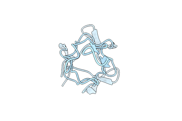
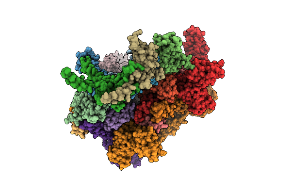
Organism: Escherichia phage p1
Method: ELECTRON MICROSCOPY Release Date: 2025-10-22 Classification: VIRAL PROTEIN |
 |
Organism: Escherichia phage p1
Method: ELECTRON MICROSCOPY Release Date: 2025-10-22 Classification: VIRAL PROTEIN |
 |
Organism: Escherichia phage p1
Method: ELECTRON MICROSCOPY Release Date: 2025-10-22 Classification: VIRAL PROTEIN |
 |
Organism: Escherichia phage p1
Method: ELECTRON MICROSCOPY Release Date: 2025-10-22 Classification: VIRAL PROTEIN |
 |
Organism: Escherichia phage p1
Method: ELECTRON MICROSCOPY Release Date: 2025-10-22 Classification: VIRAL PROTEIN |
 |
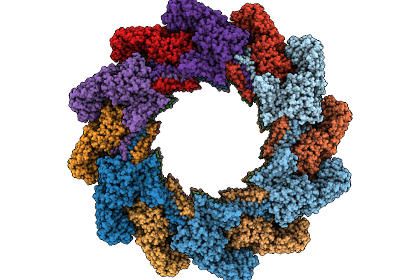
Organism: Escherichia phage p1
Method: ELECTRON MICROSCOPY Release Date: 2025-10-22 Classification: VIRAL PROTEIN |
 |
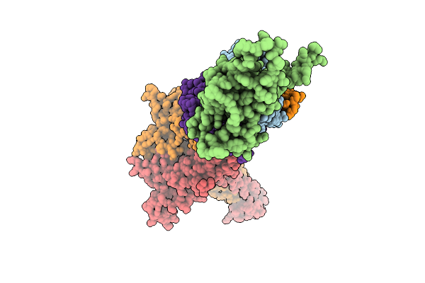
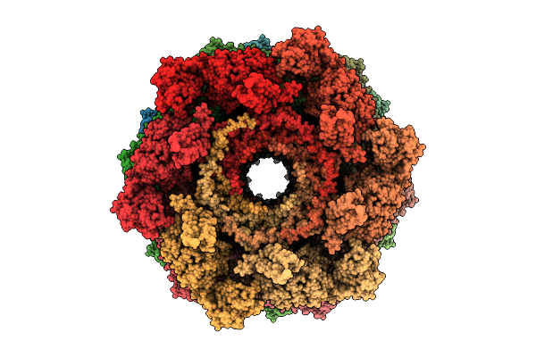
Organism: Escherichia phage p1
Method: ELECTRON MICROSCOPY Release Date: 2025-09-10 Classification: VIRAL PROTEIN |
 |
Organism: Escherichia phage p1
Method: ELECTRON MICROSCOPY Release Date: 2025-09-10 Classification: VIRAL PROTEIN |
 |
Organism: Escherichia phage p1
Method: ELECTRON MICROSCOPY Release Date: 2025-09-10 Classification: VIRAL PROTEIN |
 |
Organism: Escherichia phage p1
Method: ELECTRON MICROSCOPY Release Date: 2025-09-10 Classification: VIRAL PROTEIN |
 |
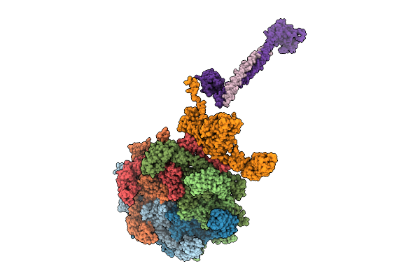
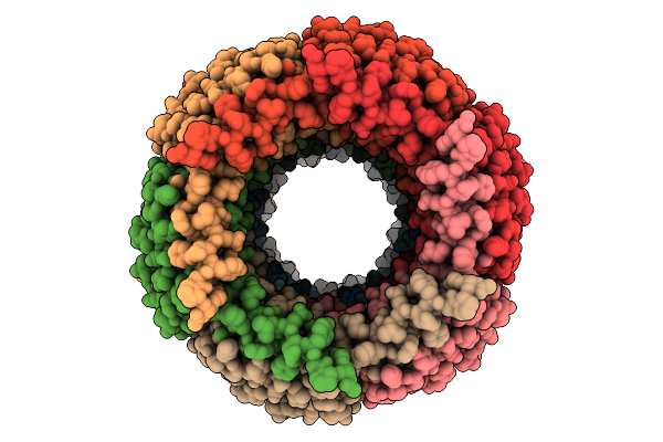
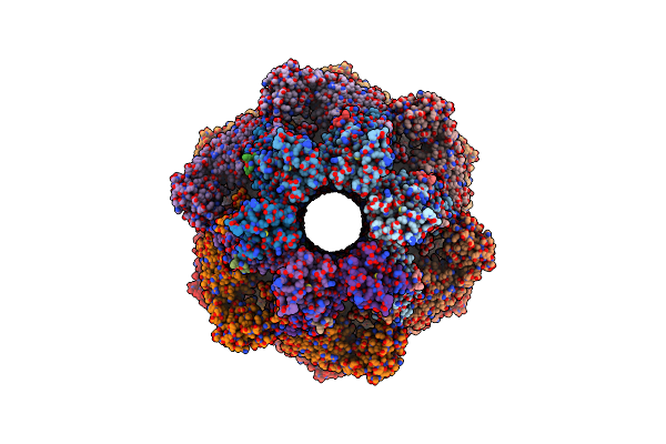
In Situ Structures Of The Ultra-Long Contracted Tail Of Myoviridae Phage P1
Organism: Escherichia phage p1
Method: ELECTRON MICROSCOPY Release Date: 2023-06-21 Classification: VIRAL PROTEIN |
 |
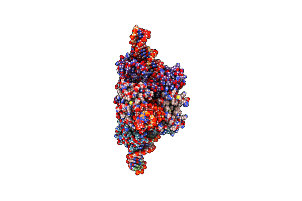
Organism: Escherichia phage p1
Method: ELECTRON MICROSCOPY Release Date: 2023-06-21 Classification: VIRAL PROTEIN |
 |
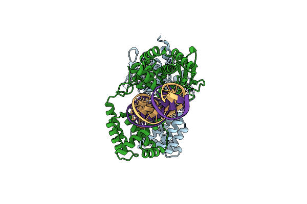
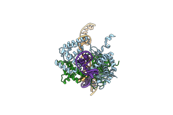
Organism: Escherichia phage p1
Method: ELECTRON MICROSCOPY Release Date: 2022-01-19 Classification: RECOMBINATION/DNA |
 |
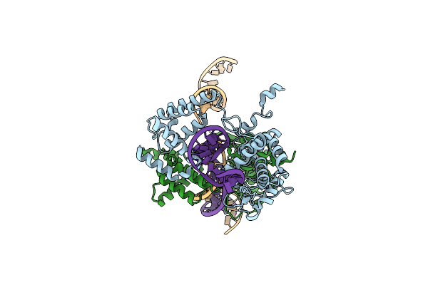
Organism: Escherichia phage p1
Method: ELECTRON MICROSCOPY Release Date: 2022-01-19 Classification: RECOMBINATION/DNA |
 |
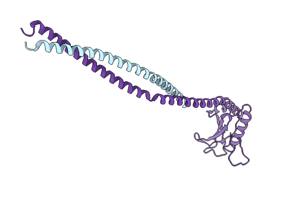
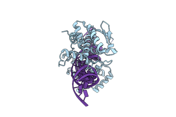
Heterodimer Of Cre Recombinase Mutants D33A/A36V/R192A And R72E/L115D/R119D In Complex With Loxp Dna.
Organism: Escherichia phage p1
Method: ELECTRON MICROSCOPY Release Date: 2022-01-19 Classification: RECOMBINATION/DNA |
 |
Basis For A Switch In Substrate Specificity: Crystal Structure Of Selected Variant Of Cre Site-Specific Recombinase, Alshg Bound To The Engineered Recognition Site Loxm7
Organism: Escherichia phage p1, Escherichia virus p1
Method: X-RAY DIFFRACTION Resolution:2.35 Å Release Date: 2004-02-17 Classification: RECOMBINATION/DNA |
 |
Basis For A Switch In Substrate Specificity: Crystal Structure Of Selected Variant Of Cre Site-Specific Recombinase, Lnsgg Bound To The Engineered Recognition Site Loxm7
Organism: Escherichia phage p1, Escherichia virus p1
Method: X-RAY DIFFRACTION Resolution:2.75 Å Release Date: 2004-02-17 Classification: RECOMBINATION/DNA |