
Search Count: 219
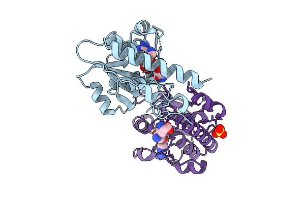 |
Organism: Escherichia coli bl21(de3)
Method: X-RAY DIFFRACTION Release Date: 2025-12-17 Classification: TRANSFERASE Ligands: SFG, ZN, SO4 |
 |
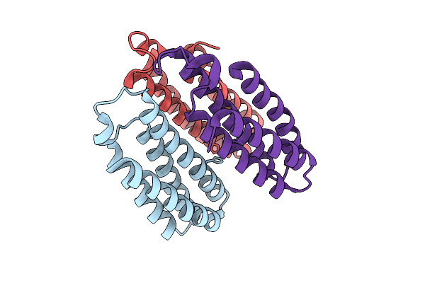
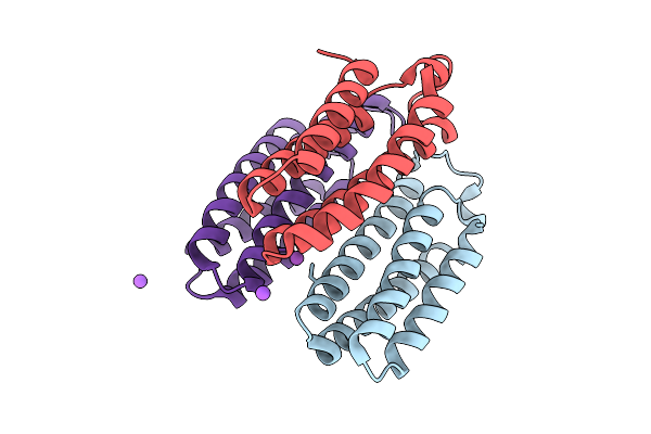
Cryo-Em Structure Of Ctpa S300A/K325A/Q329A Mutant From Helicobacter Pylori
Organism: Helicobacter pylori g27, Escherichia coli bl21(de3)
Method: ELECTRON MICROSCOPY Release Date: 2025-12-03 Classification: HYDROLASE |
 |
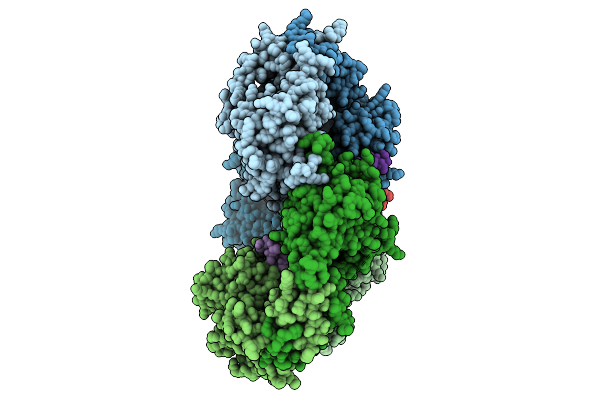
Nanomer Msp1 From S.Cerevisiae (With A Catalytic Dead Mutation) In Complex With An Unknown Peptide Substrate
Organism: Saccharomyces cerevisiae s288c, Escherichia coli bl21(de3)
Method: ELECTRON MICROSCOPY Release Date: 2025-11-26 Classification: MEMBRANE PROTEIN Ligands: ATP, MG |
 |
Organism: Escherichia coli bl21(de3)
Method: X-RAY DIFFRACTION Release Date: 2025-10-22 Classification: METAL BINDING PROTEIN Ligands: CO, CA, CL, NA |
 |
Organism: Escherichia coli bl21(de3)
Method: X-RAY DIFFRACTION Release Date: 2025-10-22 Classification: METAL BINDING PROTEIN Ligands: NI, CL, CA |
 |
Organism: Escherichia coli bl21(de3)
Method: X-RAY DIFFRACTION Release Date: 2025-10-22 Classification: METAL BINDING PROTEIN |
 |
Organism: Escherichia coli bl21(de3)
Method: X-RAY DIFFRACTION Release Date: 2025-10-22 Classification: METAL BINDING PROTEIN Ligands: CU |
 |
Organism: Escherichia coli bl21(de3)
Method: X-RAY DIFFRACTION Release Date: 2025-10-22 Classification: METAL BINDING PROTEIN Ligands: ZN, CA, NA |
 |
Organism: Escherichia coli bl21(de3)
Method: X-RAY DIFFRACTION Release Date: 2025-10-22 Classification: METAL BINDING PROTEIN Ligands: ZN, CL, BS3, NA |
 |
Organism: Escherichia coli bl21(de3)
Method: X-RAY DIFFRACTION Release Date: 2025-10-22 Classification: METAL BINDING PROTEIN Ligands: BS3, NA, CO |
 |
Organism: Escherichia coli bl21(de3)
Method: X-RAY DIFFRACTION Release Date: 2025-10-22 Classification: METAL BINDING PROTEIN Ligands: BS3, NI, CA |
 |
Organism: Escherichia coli bl21(de3)
Method: X-RAY DIFFRACTION Release Date: 2025-10-22 Classification: METAL BINDING PROTEIN Ligands: CL, NA, BS3 |
 |
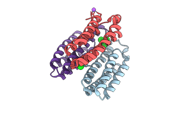
Organism: Helicobacter pylori g27, Escherichia coli bl21(de3)
Method: X-RAY DIFFRACTION Release Date: 2025-10-01 Classification: HYDROLASE |
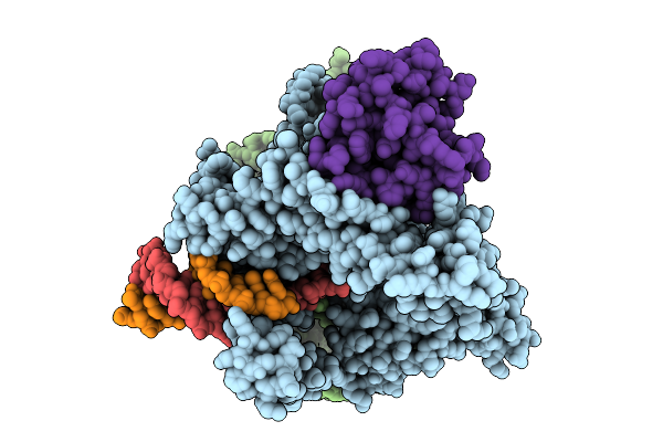 |
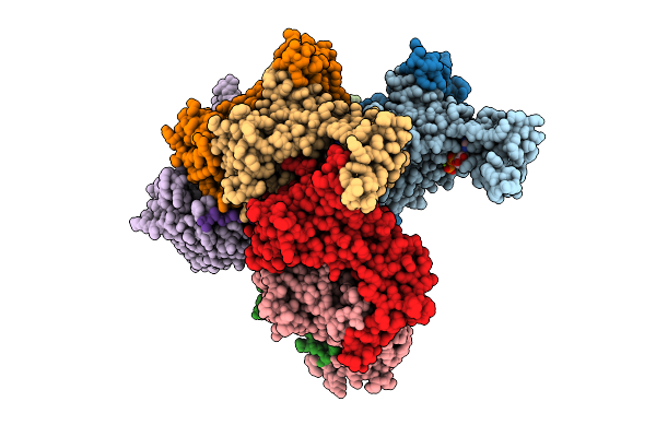
Structure Of Cas12P-Trxa-Guide Rna-Target Dna Complex(29Nt Ts And 11Nt Nts)
Organism: Unidentified, Synthetic construct, Escherichia coli bl21(de3)
Method: ELECTRON MICROSCOPY Release Date: 2025-09-10 Classification: RNA BINDING PROTEIN/RNA/DNA |
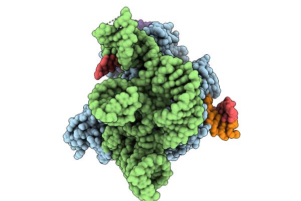 |
Organism: Unidentified, Synthetic construct, Escherichia coli bl21(de3)
Method: ELECTRON MICROSCOPY Release Date: 2025-09-10 Classification: RNA BINDING PROTEIN/RNA/DNA |
 |
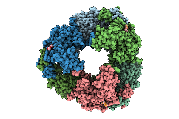
Crystal Structure Of Escherichia Coli Groel With Magnesium Ions And A Phosphorylated Serine Residue-3.2A
Organism: Escherichia coli bl21(de3)
Method: X-RAY DIFFRACTION Release Date: 2025-09-03 Classification: CHAPERONE Ligands: HEZ, MG, BME, TAM |
 |
Crystal Structure Of Escherichia Coli Groel With Magnesium Ions And A Phosphorylated Serine Residue-3.0 A
Organism: Escherichia coli bl21(de3)
Method: X-RAY DIFFRACTION Release Date: 2025-09-03 Classification: CHAPERONE Ligands: BME, HEZ, MG |
 |
Organism: Escherichia coli bl21(de3)
Method: X-RAY DIFFRACTION Release Date: 2025-08-27 Classification: ISOMERASE Ligands: QCF, PE8, SO4 |
 |
Organism: Escherichia coli bl21(de3)
Method: X-RAY DIFFRACTION Release Date: 2025-08-20 Classification: TRANSCRIPTION |
 |
Organism: Escherichia coli bl21(de3)
Method: X-RAY DIFFRACTION Release Date: 2025-08-20 Classification: HYDROLASE Ligands: CA |