
Search Count: 65
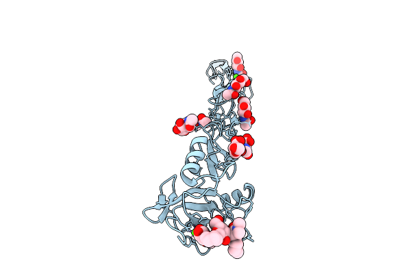 |
Organism: Homo sapiens
Method: X-RAY DIFFRACTION Release Date: 2025-12-03 Classification: CELL ADHESION Ligands: NAG, CA, A1IUQ |
 |
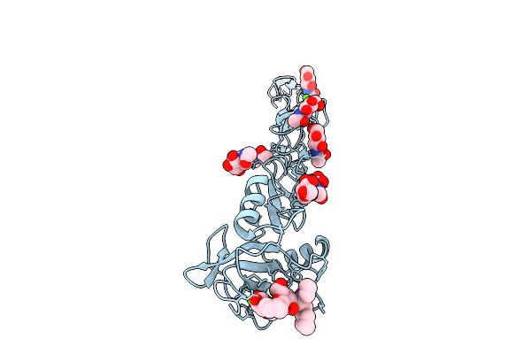
Organism: Homo sapiens
Method: X-RAY DIFFRACTION Release Date: 2025-12-03 Classification: CELL ADHESION Ligands: NAG, A1IUR, CA |
 |
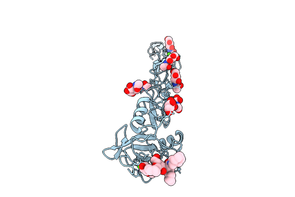
Organism: Homo sapiens
Method: X-RAY DIFFRACTION Release Date: 2025-12-03 Classification: CELL ADHESION Ligands: NAG, A1IUS, CA |
 |
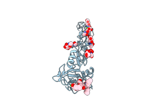
Organism: Homo sapiens
Method: X-RAY DIFFRACTION Release Date: 2025-12-03 Classification: CELL ADHESION Ligands: NAG, A1IUT, CA |
 |
Organism: Homo sapiens
Method: X-RAY DIFFRACTION Release Date: 2025-12-03 Classification: CELL ADHESION Ligands: NAG, A1IUU, CA |
 |
Organism: Homo sapiens
Method: X-RAY DIFFRACTION Resolution:1.80 Å Release Date: 2024-11-13 Classification: SUGAR BINDING PROTEIN Ligands: CA, A1H5Z |
 |
Organism: Homo sapiens
Method: X-RAY DIFFRACTION Resolution:1.90 Å Release Date: 2024-11-13 Classification: SUGAR BINDING PROTEIN Ligands: CA, A1H5Y |
 |
Organism: Homo sapiens
Method: X-RAY DIFFRACTION Resolution:2.90 Å Release Date: 2024-11-13 Classification: SUGAR BINDING PROTEIN Ligands: CA, A1H5X |
 |
Organism: Homo sapiens
Method: X-RAY DIFFRACTION Resolution:2.20 Å Release Date: 2024-10-23 Classification: CELL ADHESION Ligands: NAG, CA, Y6T |
 |
Organism: Homo sapiens
Method: X-RAY DIFFRACTION Resolution:2.49 Å Release Date: 2024-10-23 Classification: CELL ADHESION Ligands: NAG, Y6O, CA |
 |
Crystal Structure Of Dc-Sign In Complex With A Triazole-Based Glycomimetic Ligand
Organism: Homo sapiens
Method: X-RAY DIFFRACTION Resolution:2.20 Å Release Date: 2021-10-27 Classification: SUGAR BINDING PROTEIN Ligands: CA, UH8, NO3 |
 |
Crystal Structure Of Dc-Sign In Complex With A Triazole-Based Glycomimetic Ligand
Organism: Homo sapiens
Method: X-RAY DIFFRACTION Resolution:2.10 Å Release Date: 2021-10-27 Classification: SUGAR BINDING PROTEIN Ligands: CA, UH5 |
 |
Structure Of The N Terminal Domain Of Bc2L-C Lectin (1-131) In Complex With Globo H (H-Type 3) And Cas No 912569-62-1
Organism: Burkholderia cenocepacia (strain atcc baa-245 / dsm 16553 / lmg 16656 / nctc 13227 / j2315 / cf5610)
Method: X-RAY DIFFRACTION Resolution:1.90 Å Release Date: 2021-04-07 Classification: SUGAR BINDING PROTEIN Ligands: QT5, NA |
 |
Structure Of The Apo Form Of The N Terminal Domain Of Bc2L-C Lectin (1-131)
Organism: Burkholderia cenocepacia j2315
Method: X-RAY DIFFRACTION Resolution:1.50 Å Release Date: 2021-04-07 Classification: SUGAR BINDING PROTEIN |
 |
Crystal Structure Of The Lectin Domain Of The Fimh Variant Arg98Ala, In Complex With Methyl 3-Chloro-4-D-Mannopyranosyloxy-3-Biphenylcarboxylate
Organism: Escherichia coli k12
Method: X-RAY DIFFRACTION Resolution:1.42 Å Release Date: 2020-12-23 Classification: CELL ADHESION Ligands: SBQ, EDO |
 |
Crystal Structure Of Fimh In Complex With A Tetraflourinated Biphenyl Alpha D-Mannoside
Organism: Escherichia coli (strain k12)
Method: X-RAY DIFFRACTION Resolution:2.10 Å Release Date: 2019-03-20 Classification: CELL ADHESION Ligands: EJK, SO4, EPE |
 |
Crystal Structure Of Fimh In Complex With A Pentaflourinated Biphenyl Alpha D-Mannoside
Organism: Escherichia coli (strain k12)
Method: X-RAY DIFFRACTION Resolution:2.20 Å Release Date: 2019-03-20 Classification: CELL ADHESION Ligands: EJN, SO4 |
 |
Crystal Structure Of A Fimh*Dsg Complex From E.Coli F18 With Bound Trimannose
Organism: Escherichia coli f18+, Escherichia coli 536
Method: X-RAY DIFFRACTION Resolution:2.10 Å Release Date: 2019-01-16 Classification: CELL ADHESION Ligands: PEG, 1PE |
 |
Crystal Structure Of The Fimh Lectin Domain From E.Coli F18 In Complex With Trimannose
Organism: Escherichia coli f18+
Method: X-RAY DIFFRACTION Resolution:2.50 Å Release Date: 2019-01-16 Classification: CELL ADHESION Ligands: CA |
 |
Crystal Structure Of The Fimh Lectin Domain From E.Coli K12 In Complex With The Dimannoside Man(Alpha1-2)Man
Organism: Escherichia coli (strain k12)
Method: X-RAY DIFFRACTION Resolution:2.50 Å Release Date: 2019-01-16 Classification: CELL ADHESION Ligands: SO4 |